What Constitutes a True Lateral Wrist Position?
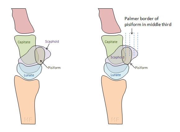
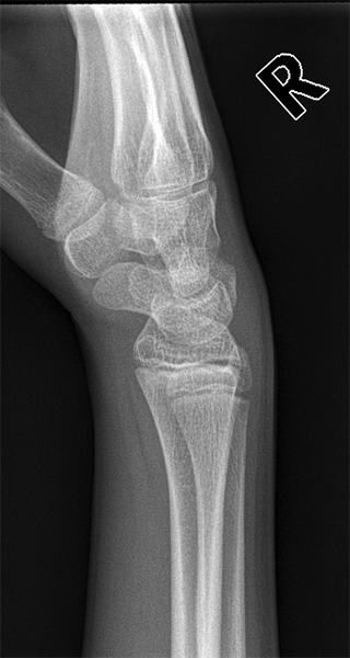
Definition of a Lateral Wrist- The radius and Carpal BonesThe term true lateral frequently appears on request forms for plain film X-ray examinations. Can the term, when applied to the wrist, be defined with more precision than a vague notion of best lateralness? This page considers a definition of a true lateral wrist that goes beyond the mere consideration of whether the radius and ulna overlap.
drawing based on
<a class="external" href="http://radiology.rsnajnls.org/cgi/content/full/219/1/11" rel="nofollow" target="_blank">Charles A. Goldfarb, MD, Yuming Yin, MD, Louis A. Gilula, MD, Andrew J. Fisher, MD and Martin I. Boyer, MD
</a><a class="external" href="http://radiology.rsnajnls.org/cgi/content/full/219/1/11" rel="nofollow" target="_blank">What the Clinician Wants to Know</a>
<a class="external" href="http://radiology.rsnajnls.org/cgi/content/full/219/1/11" rel="nofollow" target="_blank"> Wrist Fractures: What the Clinician Wants to Know1 </a>
<a class="external" href="http://radiology.rsnajnls.org/cgi/content/full/219/1/11" rel="nofollow" target="_blank"> Radiology. 2001;219:11-28.</a>
quoted from <a class="external" href="http://www.radiologyassistant.nl/en/476a23436683b" rel="nofollow" target="_blank">http://www.radiologyassistant.nl/en/476a23436683b</a>
adapted from
<a class="external" href="http://radiology.rsnajnls.org/cgi/content/full/219/1/11" rel="nofollow" target="_blank">Charles A. Goldfarb, MD, Yuming Yin, MD, Louis A. Gilula, MD, Andrew J. Fisher, MD and Martin I. Boyer, MD
</a><a class="external" href="http://radiology.rsnajnls.org/cgi/content/full/219/1/11" rel="nofollow" target="_blank">What the Clinician Wants to Know</a>
<a class="external" href="http://radiology.rsnajnls.org/cgi/content/full/219/1/11" rel="nofollow" target="_blank"> Wrist Fractures: What the Clinician Wants to Know1 </a>
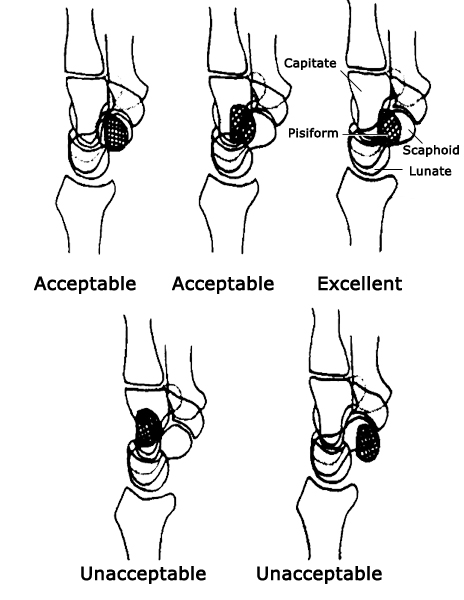
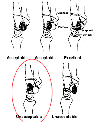
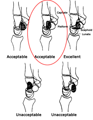
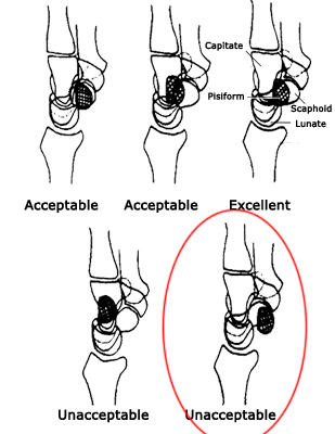
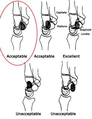
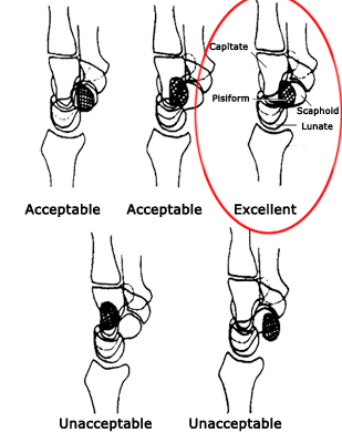
<a class="external" href="http://radiology.rsnajnls.org/cgi/content/full/219/1/11" rel="nofollow" target="_blank"> Radiology. 2001;219:11-28.</a>These authors go one step further by establishing the bounds of acceptability of lateral wrist positioning.
Examples
adapted from
<a class="external" href="http://radiology.rsnajnls.org/cgi/content/full/219/1/11" rel="nofollow" target="_blank">Charles A. Goldfarb, MD, Yuming Yin, MD, Louis A. Gilula, MD, Andrew J. Fisher, MD and Martin I. Boyer, MD
</a><a class="external" href="http://radiology.rsnajnls.org/cgi/content/full/219/1/11" rel="nofollow" target="_blank">What the Clinician Wants to Know</a>
<a class="external" href="http://radiology.rsnajnls.org/cgi/content/full/219/1/11" rel="nofollow" target="_blank"> Wrist Fractures: What the Clinician Wants to Know1 </a>
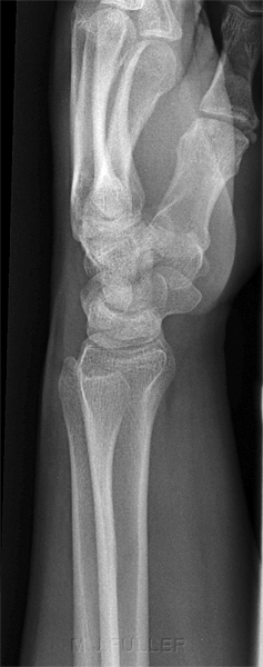
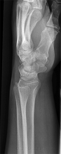
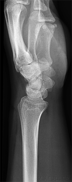
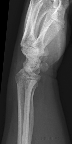
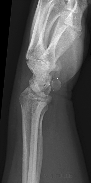
<a class="external" href="http://radiology.rsnajnls.org/cgi/content/full/219/1/11" rel="nofollow" target="_blank"> Radiology. 2001;219:11-28.</a>The wrist is malpositioned- too pronated pisiform outlined This position is unacceptable using the Goldfarb et al criteria
adapted from
<a class="external" href="http://radiology.rsnajnls.org/cgi/content/full/219/1/11" rel="nofollow" target="_blank">Charles A. Goldfarb, MD, Yuming Yin, MD, Louis A. Gilula, MD, Andrew J. Fisher, MD and Martin I. Boyer, MD
</a><a class="external" href="http://radiology.rsnajnls.org/cgi/content/full/219/1/11" rel="nofollow" target="_blank">What the Clinician Wants to Know</a>
<a class="external" href="http://radiology.rsnajnls.org/cgi/content/full/219/1/11" rel="nofollow" target="_blank"> Wrist Fractures: What the Clinician Wants to Know1 </a>
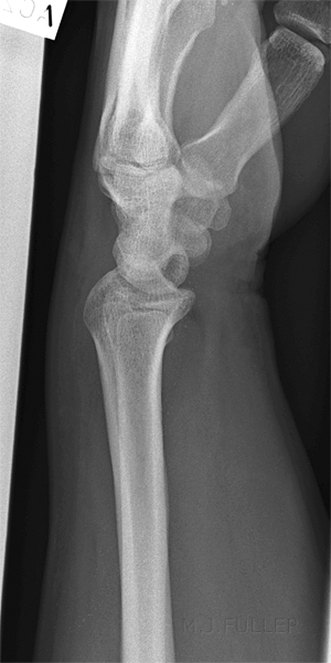
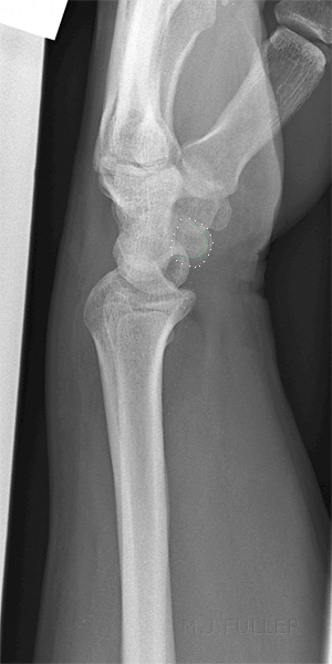
<a class="external" href="http://radiology.rsnajnls.org/cgi/content/full/219/1/11" rel="nofollow" target="_blank"> Radiology. 2001;219:11-28.</a>An apparently well positioned lateral wrist! pisiform outlined This position is acceptable using the Goldfarb et al criteria. The palmer surface of the pisiform appears to fall within the more dorsal third of the acceptability criteria regions
adapted from
<a class="external" href="http://radiology.rsnajnls.org/cgi/content/full/219/1/11" rel="nofollow" target="_blank">Charles A. Goldfarb, MD, Yuming Yin, MD, Louis A. Gilula, MD, Andrew J. Fisher, MD and Martin I. Boyer, MD
</a><a class="external" href="http://radiology.rsnajnls.org/cgi/content/full/219/1/11" rel="nofollow" target="_blank">What the Clinician Wants to Know</a>
<a class="external" href="http://radiology.rsnajnls.org/cgi/content/full/219/1/11" rel="nofollow" target="_blank"> Wrist Fractures: What the Clinician Wants to Know1 </a>
<a class="external" href="http://radiology.rsnajnls.org/cgi/content/full/219/1/11" rel="nofollow" target="_blank"> Radiology. 2001;219:11-28.</a>A prima facie well positioned lateral wrist! pisiform outlined This position is unacceptable using the Goldfarb et al criteria. The palmer surface of the pisiform appears to fall more palmarly than the scaphoid. It is possible that the criteria have failed in this case because the patient had sustained a scaphoid fracture previously. The lunate is also noted to be dorsally tilted.
adapted from
<a class="external" href="http://radiology.rsnajnls.org/cgi/content/full/219/1/11" rel="nofollow" target="_blank">Charles A. Goldfarb, MD, Yuming Yin, MD, Louis A. Gilula, MD, Andrew J. Fisher, MD and Martin I. Boyer, MD
</a><a class="external" href="http://radiology.rsnajnls.org/cgi/content/full/219/1/11" rel="nofollow" target="_blank">What the Clinician Wants to Know</a>
<a class="external" href="http://radiology.rsnajnls.org/cgi/content/full/219/1/11" rel="nofollow" target="_blank"> Wrist Fractures: What the Clinician Wants to Know1 </a>
<a class="external" href="http://radiology.rsnajnls.org/cgi/content/full/219/1/11" rel="nofollow" target="_blank"> Radiology. 2001;219:11-28.</a>Appears to be lateral pisiform outlined This position is acceptable using the Goldfarb et al criteria...but only just. The palmer surface of the pisiform is only just more dorsally located than the palmer surface of the scaphoid.
adapted from
<a class="external" href="http://radiology.rsnajnls.org/cgi/content/full/219/1/11" rel="nofollow" target="_blank">Charles A. Goldfarb, MD, Yuming Yin, MD, Louis A. Gilula, MD, Andrew J. Fisher, MD and Martin I. Boyer, MD
</a><a class="external" href="http://radiology.rsnajnls.org/cgi/content/full/219/1/11" rel="nofollow" target="_blank">What the Clinician Wants to Know</a>
<a class="external" href="http://radiology.rsnajnls.org/cgi/content/full/219/1/11" rel="nofollow" target="_blank"> Wrist Fractures: What the Clinician Wants to Know1 </a>
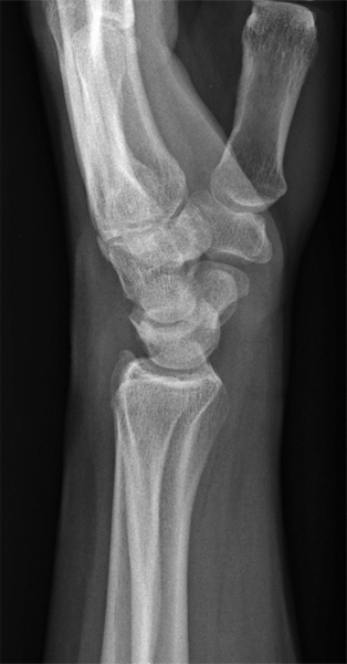
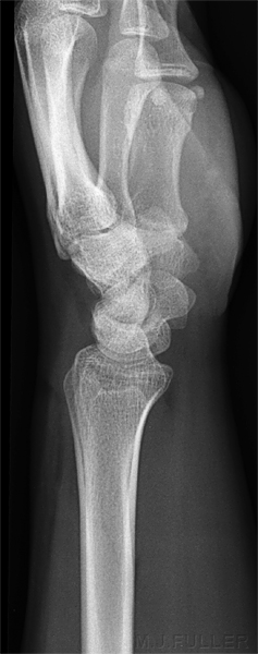
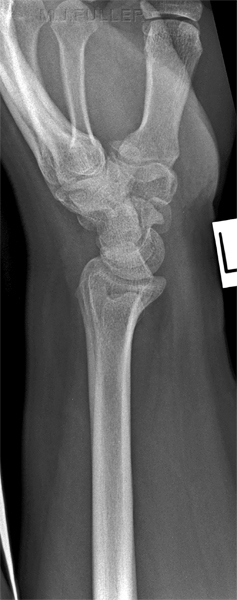
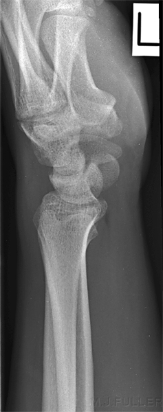
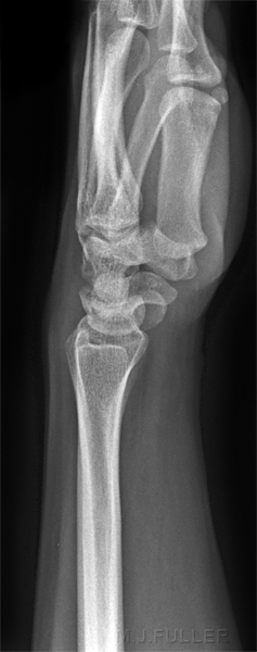
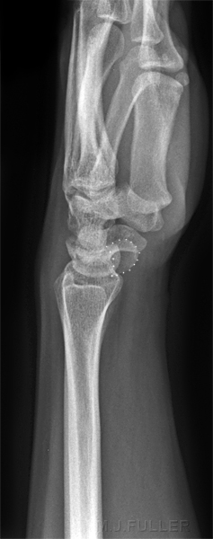
<a class="external" href="http://radiology.rsnajnls.org/cgi/content/full/219/1/11" rel="nofollow" target="_blank"> Radiology. 2001;219:11-28.</a>This lateral wrist is dorsally rotated (rolled too far back). The pisiform (dotted) is almost projected clear of the carpal bones. This is a fail. Note however the difference in the appearance of the scaphoid compared to the cased above. Are these lateral wrists passing and failing depending on the shape of the patient's scaphoid? (..to some degree)
adapted from
<a class="external" href="http://radiology.rsnajnls.org/cgi/content/full/219/1/11" rel="nofollow" target="_blank">Charles A. Goldfarb, MD, Yuming Yin, MD, Louis A. Gilula, MD, Andrew J. Fisher, MD and Martin I. Boyer, MD
</a><a class="external" href="http://radiology.rsnajnls.org/cgi/content/full/219/1/11" rel="nofollow" target="_blank">What the Clinician Wants to Know</a>
<a class="external" href="http://radiology.rsnajnls.org/cgi/content/full/219/1/11" rel="nofollow" target="_blank"> Wrist Fractures: What the Clinician Wants to Know1 </a>
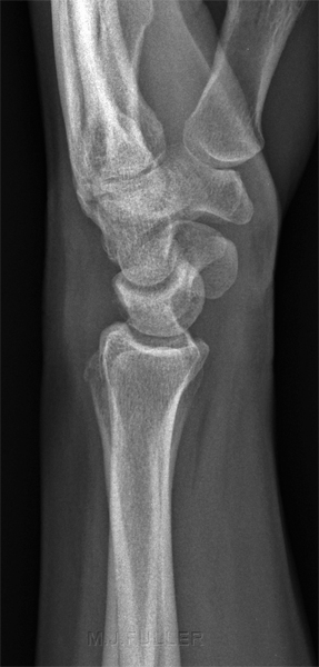
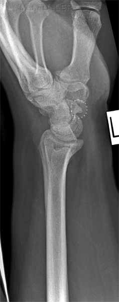
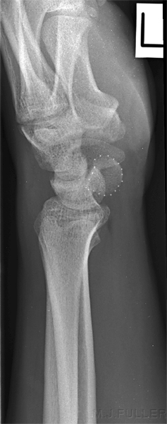
<a class="external" href="http://radiology.rsnajnls.org/cgi/content/full/219/1/11" rel="nofollow" target="_blank"> Radiology. 2001;219:11-28.</a>Same patient as above. The radiographer has recognised the positioning error and rotated the patient's wrist in a palmar (volar) direction. The pisiform (dotted white) is now projected over the distal pole of the scaphoid (dotted green). This is an improved position but still needs to be rotated further in a palmar direction to achieve a true lateral.
If you look at the malpositioned wrist above, the carpal bone anatomy is more difficult to identify than in this more correctly positioned repeat view.Despite the improved lateralness, this position would still be considered a failed lateral according to the Goldfarb et al criteria.
adapted from
<a class="external" href="http://radiology.rsnajnls.org/cgi/content/full/219/1/11" rel="nofollow" target="_blank">Charles A. Goldfarb, MD, Yuming Yin, MD, Louis A. Gilula, MD, Andrew J. Fisher, MD and Martin I. Boyer, MD
</a><a class="external" href="http://radiology.rsnajnls.org/cgi/content/full/219/1/11" rel="nofollow" target="_blank">What the Clinician Wants to Know</a>
<a class="external" href="http://radiology.rsnajnls.org/cgi/content/full/219/1/11" rel="nofollow" target="_blank"> Wrist Fractures: What the Clinician Wants to Know1 </a>
<a class="external" href="http://radiology.rsnajnls.org/cgi/content/full/219/1/11" rel="nofollow" target="_blank"> Radiology. 2001;219:11-28.</a>Bingo- whilst not perfect, it is just within the middle third of the acceptability zone
Why is a True Lateral Wrist Position Important?
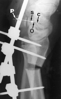
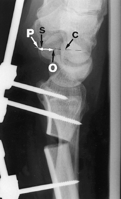
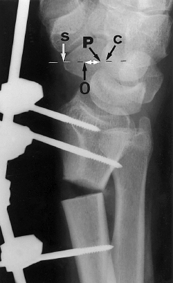
Treatment decisions for Colles fractures are partly based on the degree of angulation of the wrist associated with the fracture (palmar inclination). A poorly positioned lateral wrist will give a false indication of the degree of palmar inclination. The images shown below are of a cadaver experiment in which a fracture of known angulation (20 degrees) was imaged in various lateral and off-lateral positions. In this study, the authors demonstrated that supination increased the apparent palmar tilt of the distal radius. The authors reported that as the distance 'P' incremented by 1mm, so did the apparent angular displacement of the fracture. This makes for a simple rule of thumb to calculate how much an off-lateral wrist image is affecting the apparent angular displacement of the fracture. The take home message here is that a poorly positioned lateral wrist will potentially overstate or understate the degree of angular displacement of the wrist fracture. This could have an adverse effect on treatment decisions.
(a) Lateral radiograph shows supination of 20° and palmar tilt of -13°.
<a class="external" href="http://radiology.rsnajnls.org/cgi/content/full/220/3/594" rel="nofollow" target="_blank">Marco Zanetti, MD. Louis A. Gilula, MD. Hilaire A. C. Jacob, PhD. Juerg Hodler, MD Palmar Tilt of the Distal Radius: Influence of Off-lateral Projection— Initial Observations. Radiology. 2001;220:594-600.</a>(b) Lateral
radiograph shows neutral position of 0° and palmar tilt of -15°.
<a class="external" href="http://radiology.rsnajnls.org/cgi/content/full/220/3/594" rel="nofollow" target="_blank">Marco Zanetti, MD. Louis A. Gilula, MD. Hilaire A. C. Jacob, PhD. Juerg Hodler, MD Palmar Tilt of the Distal Radius: Influence of Off-lateral Projection— Initial Observations. Radiology. 2001;220:594-600.</a>(c) Radiograph shows pronation of 20° and palmar tilt of -29°.
<a class="external" href="http://radiology.rsnajnls.org/cgi/content/full/220/3/594" rel="nofollow" target="_blank">Marco Zanetti, MD. Louis A. Gilula, MD. Hilaire A. C. Jacob, PhD. Juerg Hodler, MD Palmar Tilt of the Distal Radius: Influence of Off-lateral Projection— Initial Observations. Radiology. 2001;220:594-600.</a>
Definition of a Lateral Wrist- The Ulna
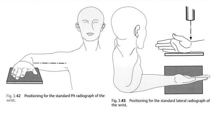
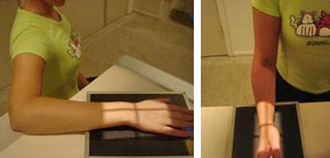
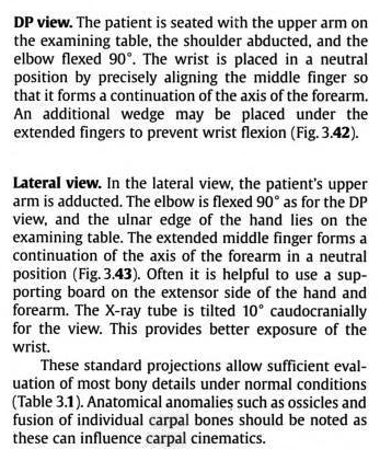
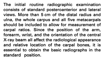
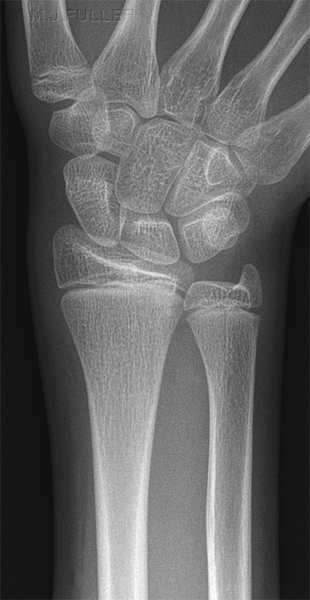
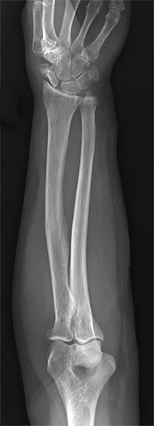
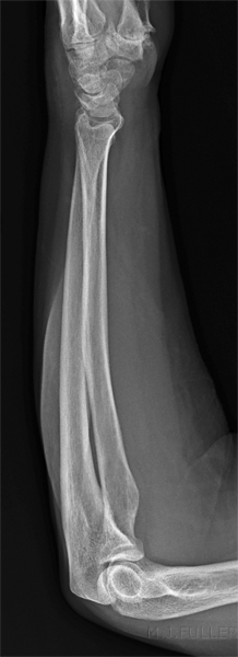
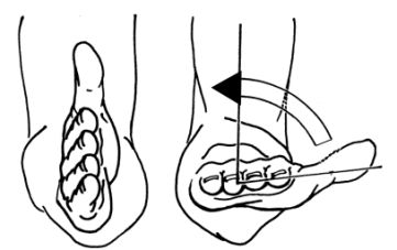
The radiographic technique advice on wrist positioning is confusing. The technique described below appears to be a Radiologist's approach to achieving "standard projections" of the wrist. Should this be considered a special technique and, if it is, when should it be applied? If it is the Radiologist's technique of choice, why shouldn't it be applied as a routine. The radiographic technique for AP and lateral wrist (as per K.C. Clarke and Merrill's atlas) is to have the patient's humerus abducted 90 degrees and the elbow flexed to 90 degrees as shown in figure 3.42 below- you simply roll (pronate/supinate) the wrist from PA to lateral without moving the elbow from the lateral position. Importantly, no mention is made of the fact that the radius rolls around the ulna resulting in a single view of the ulna. In the words of John Harris et al (The Radiology of Emergency Medicine, 3rd Ed., 1993, p375) "one view is no view". It is inevitable that ulna fractures will be missed using this technique. In the presence of a fractured radius, a missed ulna fracture could be significant in terms of treatment. A further point of confusion is that the Radiography standard positioning for forearm radiography achieves imaging of the ulna in two projections at 90 degrees. The Radiologist is thus provided with true AP/PA and lateral positioning of the wrist with forearm radiography but not with wrist radiography. Unfortunately, because of the divergent X-ray beam, the images of the wrist obtained with standard forearm radiography are of limited utility.
Comment
These descriptions are from radiology-based texts rather than radiography texts. The positions as described above are akin to 'mixed forearm' views which are performed when patients have limited movement in their arm (usually from pain) or when the patient has an above elbow cast. The wrist positioning approach described above will tend to produce two views of the anatomy at 90 degrees because the relationship between the radius and ulna does not change- the 90 degree movement occurs largely at the shoulder.
adapted from
<a class="external" href="http://books.google.com.au/books?id=G1gWHR1_J9UC&pg=PA54&lpg=PA54&dq=wrist+pronation+supination&source=bl&ots=b7aEg3_V3A&sig=cOaOxDQ3blhQsbSAY_mz-70mSR8&hl=en&ei=BIUaSs7eE4-IkAXrkj0&sa=X&oi=book_result&ct=result&resnum=5#PPA55,M1" rel="nofollow" target="_blank">Raoul Tubiana, Jean-Michel Thomine, Evelyn Mackin
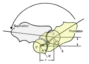
</a><a>Examination of the Hand and Wrist. Edition: 2, 1998</a>It seems counter-intuitive to suggest that when you pronate and supinate your hand, the distal ulna does not follow the movement of the radius and also rotate through 180 degrees. In fact the distal ulna moves very little during pronation/supination of the wrist. The ulna translates through an arc of a circle but does not rotate. The radius undergoes a rotation of almost 180 degrees from full pronation to supination. During this movement of the radius, the ulna translates through a short arc of a circle. Note the position of the ulna styloid.
Pronation and Supination Videos
<embed allowfullscreen="true" height="350" src="http://widget.wetpaintserv.us/wiki/wikiradiography/widget/youtubevideo/93a2833b88a7622ae4af1f33b3c54a7cbc2b796b" type="application/x-shockwave-flash" width="425" wmode="transparent"/>
adapted from
<a class="external" href="http://books.google.com.au/books?id=G1gWHR1_J9UC&pg=PA54&lpg=PA54&dq=wrist+pronation+supination&source=bl&ots=b7aEg3_V3A&sig=cOaOxDQ3blhQsbSAY_mz-70mSR8&hl=en&ei=BIUaSs7eE4-IkAXrkj0&sa=X&oi=book_result&ct=result&resnum=5#PPA55,M1" rel="nofollow" target="_blank">Raoul Tubiana, Jean-Michel Thomine, Evelyn Mackin
</a><a>Examination of the Hand and Wrist. Edition: 2, 1998</a><embed allowfullscreen="true" height="350" src="http://widget.wetpaintserv.us/wiki/wikiradiography/widget/youtubevideo/8492298a86d00ab8a8870bde3a361738d6662583" type="application/x-shockwave-flash" width="425" wmode="transparent"/> This video shows the movement of the distal radius and ulna during the movement of the wrist from PA to lateral. If you watch the ulna (and ulnar styloid), there is no rotation movement.
(Note: Youtube videos are displayed at lower resolution when 'hot-linked'. Click on the bottom right corner of the video and it will open at full resolution.)This video may assist in your understanding of the pronation/supination rotation of the wrist
Discussion
Using the criteria described above a radiographer can determine with accuracy the acceptability of a lateral wrist image. When the patient has suffered a wrist fracture and the palmar tilt is likely to be pivotal in treatment decisions, the radiographer can make an informed judgement as to whether to repeat the view.
... back to the Applied Radiography home page
... back to the Wikiradiography home page
