Triquetral Fractures
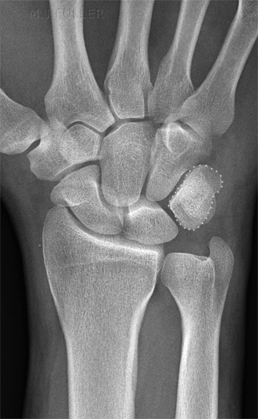
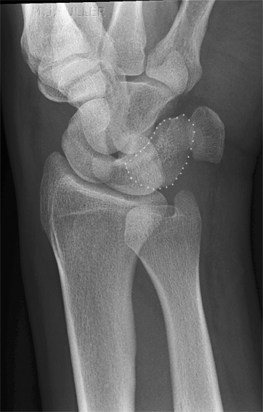
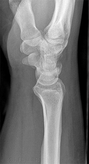
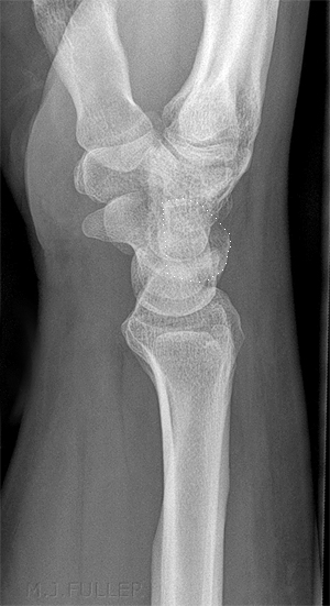
Triquetral fractures are the second most common fracture of the carpal bones. They can easily be missed. A knowledge of mechanism of injury, anatomy and radiographic technique can be useful for the radiographer in demonstrating these fractures.
Anatomy
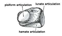
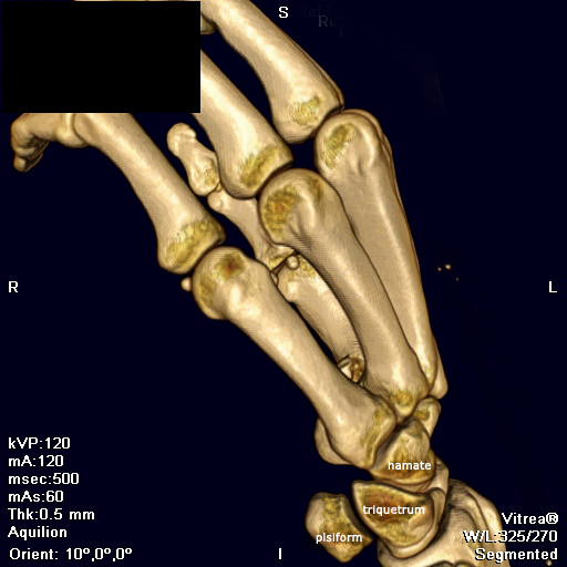
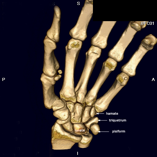
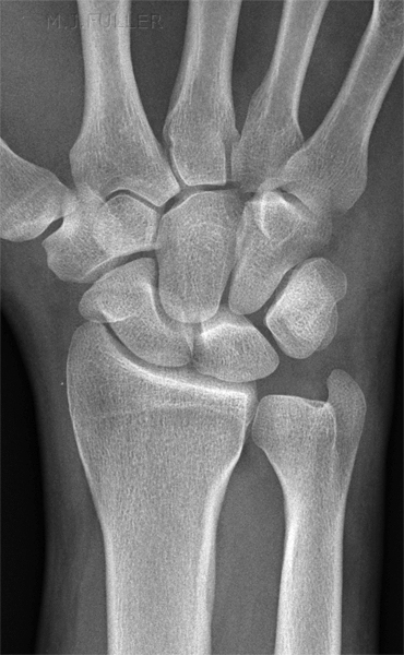
<a class="external" href="http://education.yahoo.com/reference/gray/subjects/subject/54#p224" rel="nofollow" target="_blank">adapted from Gray's anatomy</a>The triquetrum may be distinguished by its pyramidal shape, and by an oval isolated facet for articulation with the pisiform bone. It is situated at the upper and ulnar side of the carpus. The superior surface presents a medial, rough, non-articular portion, and a lateral convex articular portion which articulates with the triangular articular disk of the wrist. The inferior surface, directed lateralward, is concave, sinuously curved, and smooth for articulation with the hamate. The dorsal surface is rough for the attachment of ligaments. The volar surface presents, on its medial part, an oval facet, for articulation with the pisiform; its lateral part is rough for ligamentous attachment. The lateral surface, the base of the pyramid, is marked by a flat, quadrilateral facet, for articulation with the lunate. The medial surface, the summit of the pyramid, is pointed and roughened, for the attachment of the ulnar collateral ligament of the wrist. ( <a class="external" href="http://education.yahoo.com/reference/gray/subjects/subject/54#p224" rel="nofollow" target="_blank">http://education.yahoo.com/reference/gray/subjects/subject/54#p224</a>) This 3D CT reconstruction demonstrates the spatial relationship between the triquetrum and the pisiform, hamate and lunate (not marked, largely obscured by triquetrum). This 3D CT reconstruction demonstrates the spatial relationship between the triquetrum and the pisiform, hamate and lunate .
Radiography
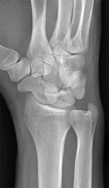
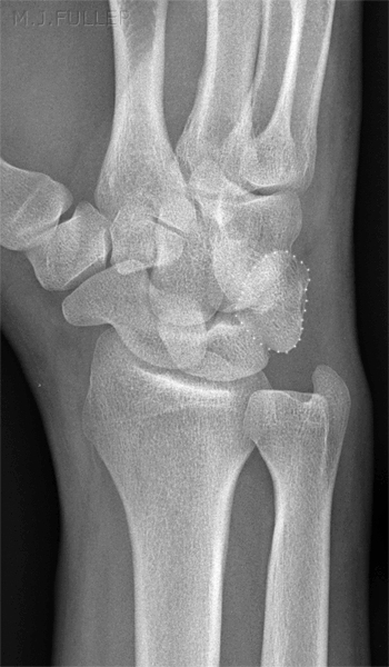
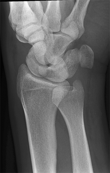
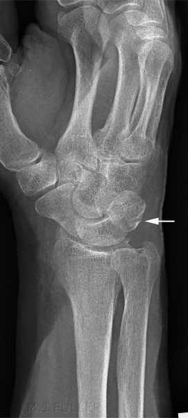
An off-lateral can be a very useful supplementary view. This is a minimally obliqued lateral wrist position. Where a triquetral fracture is suspected, both off-lateral positions may be of assistance in positively identifying the fracture. What is most likely achieved with an off-lateral position is a tangential view of the avulsion fracture. The reverse oblique is particularly useful in demonstrating the articulation between the triquetrum and the pisiform.
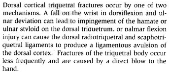
Mechanism of Injury
<a class="external" href="http://books.google.com.au/books?id=O2J1MnhH0LoC&pg=RA2-PA480&lpg=RA2-PA480&dq=triquetral+fracture+mechanism&source=bl&ots=pVcDxp0z-W&sig=sbbL66cQKniAsRoZaFGl5gx6jKg&hl=en&ei=bgLcSduLEZWVkAXv-e2iDg&sa=X&oi=book_result&ct=result&resnum=4" rel="nofollow" target="_blank">Greenberg's text-atlas of emergency medicine</a>
<a class="external" href="http://books.google.com.au/books?id=O2J1MnhH0LoC&pg=RA2-PA480&lpg=RA2-PA480&dq=triquetral+fracture+mechanism&source=bl&ots=pVcDxp0z-W&sig=sbbL66cQKniAsRoZaFGl5gx6jKg&hl=en&ei=bgLcSduLEZWVkAXv-e2iDg&sa=X&oi=book_result&ct=result&resnum=4" rel="nofollow" target="_blank">By Michael I. Greenberg, Mark Silverberg, Robert G. Hendrickson, Colleen J. Campbell. p 480</a>
Case 1
Case 2
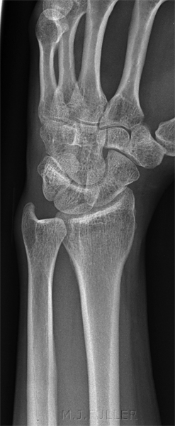
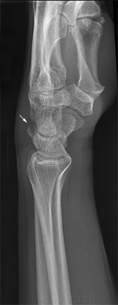
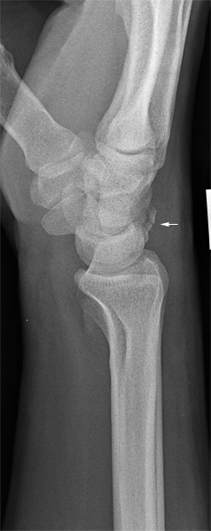
This 45 year old lady presented to the Emergency Department with a sore wrist after falling down a flight of stairs.
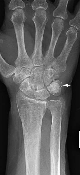
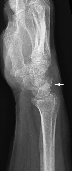
The PA image does not reveal any specific pathology Similarly, the oblique image demonstrates no displaced fracture The lateral image demonstrates a minimally displaced triquetral fracture (arrowed)
Case 3
This 38 year old man presented to the Emergency Department with a sore wrist after falling off a skateboard.
The PA image does not reveal any specific pathology Similarly, the oblique image demonstrates no displaced fracture The lateral image demonstrates a minimally displaced triquetral fracture (arrowed)
Case 4
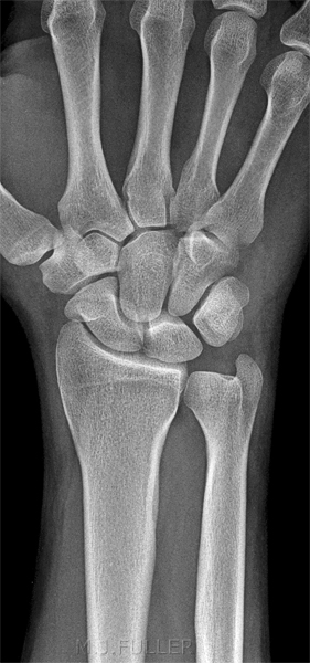
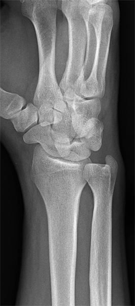
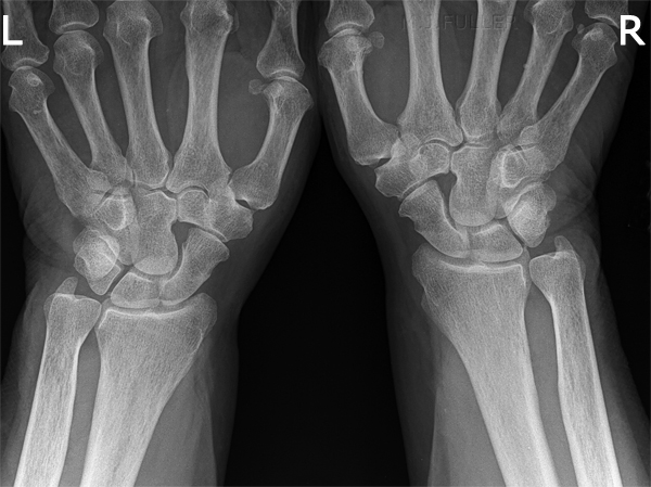
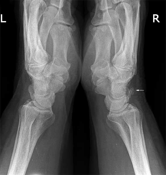
This 75 year old man presented to the Emergency Department with a sore wrist of unknown cause.
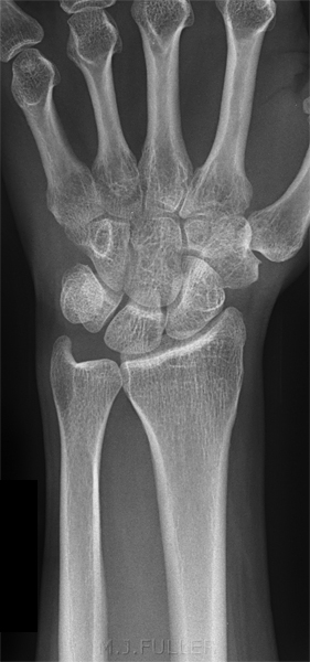
The PA image demonstrates no convincing acute injury. The lateral wrists were exposed together with the wrists in a prayer position. There is a corticated bony fragment demonstrated on the dorsum of the right wrist (arrowed) which is likely to be an old triquetrum fracture.
Harold Schubert, MD notes
"Chip fractures of the dorsal triquetrum are nonarticular; the chip might unite with the triquetrum body or, if the gap between them is 2 mm or more, a separate ossicle might form."
<a class="external" href="http://www.cfpc.ca/cfp/2000/VOL46-2000-PDFs/JAN00+PDFS/vol46-jan00-clinchal-4.pdf" rel="nofollow" target="_blank">Harold Schubert, Emergency Case Triquetrum fracture</a>
...back to the Applied Radiography home page
