Thorax
Jump to navigation
Jump to search
ANTERIOR CHEST
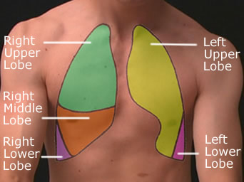
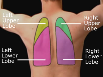
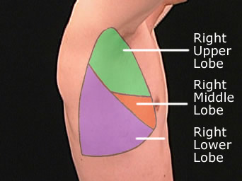
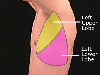
The Right Lung Consists of 3 lobes - Upper, Middle and Lower. The Left lung consists of 2 lobes - Upper and Lower.
Go Back to Surface Anatomy Index
Thorax Surface Anatomy | Other pages of interest |
Surface Anatomy Thorax
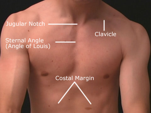
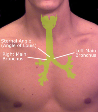
| | The jugular notch, also known as the suprasternal notch is the large, visible dip where the clavicles joins the sternum. The sternal angle is the angle formed by the junction of the manubrium and the body of the sternum in the form of a secondary cartilaginous joint (symphysis). This is also called the manubriosternal joint or Angle of Louis. This palpable clinical landmark marks the approximate level of the 2nd pair of costal cartilages and the level of the intervertebral disc between T4 and T5. It also marks approximately the beginning and end of the aortic arch and the bifurcation of the trachea into the left and right main bronchi. The clavicle or in laymans terms "collar bone" is classified as a long bone that makes up part of the shoulder girdle. The Costal margin is the lower edge of the chest (thorax) formed by the bottom edge of the rib cage. |
The Heart
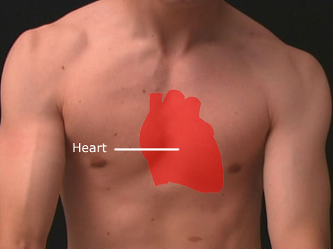
| | The heart is a muscular organ responsible for pumping blood through the blood vessels by repeated, rhythmic contractions |
Lungs
The Right Lung Consists of 3 lobes - Upper, Middle and Lower. The Left lung consists of 2 lobes - Upper and Lower.
Lungs - Anterior Projection Right Lung - Lateral
Trachea
Trachea is the name of the airway through which respiratory air moves in and out of the lungs. It bifurcates at the Sternal Angle into the Right and Left main bronchus.
Go Back to Surface Anatomy Index
