The Trauma Lateral Cervical Spine
AnatomyThe lateral cervical spine projection is one of the mainstays of trauma radiography. Its importance in the emergency room cannot be overstated. It is also a projection that fails to varying degrees. This page looks at what constitutes a good lateral cervical spine image in a trauma setting, and what can go wrong.
Getting Near the Patient
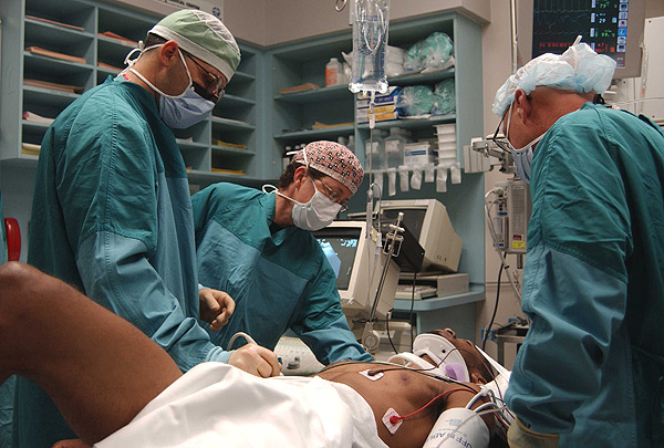
One of the first difficulties in the resus room can be getting anywhere near the patient. The more serious the trauma, the more likely the room will be full of doctors and nurses. If the patient is surrounded by staff, it is prudent to see what they are doing and make a judgement about whether your imaging is more important than what they are doing. e.g. if they are intubating the patient, stand clear..... if they are cutting off clothes, cleaning a superficial wound etc push in. Treat your role as being part of a team. Team players take into account what the other players are doing.
While you are waiting, make sure you are prepared- use the time wisely. For example, set an approximate exposure, hand out lead gowns, collect additional cassettes and other equipment, call for radiographic backup etc.
<a class="external" href="http://www.armymedicine.army.mil/news/photos/fullsize/lg_ultrasound.jpg" rel="nofollow" target="_blank">http://www.armymedicine.army.mil/news/photos/fullsize/lg_ultrasound.jpg</a>
Removing Potential Artifacts
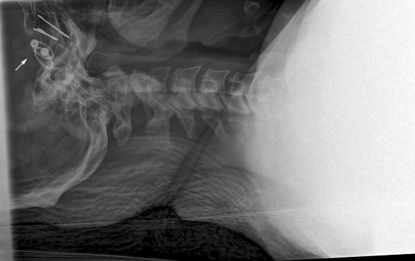
Dealing with Earrings
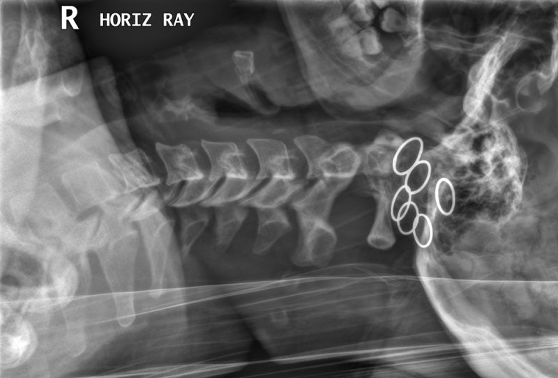
Dealing with Earrings
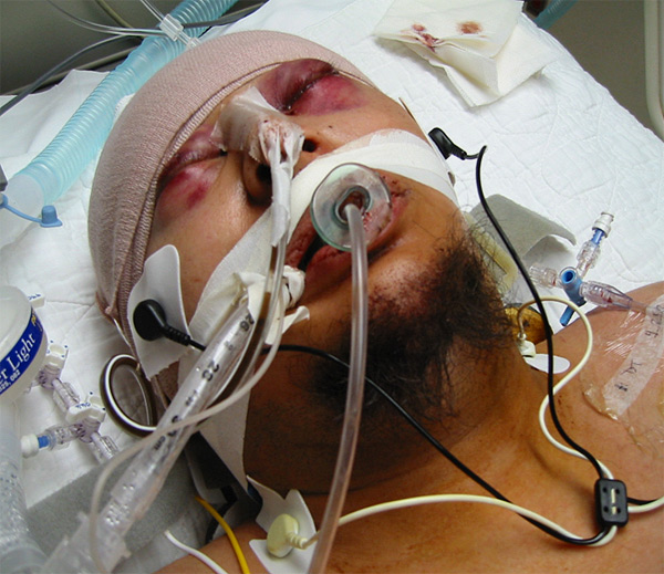
This patients earrings were hidden under the hard cervical colar. It is necessary to ask the patient if he/she is wearing earrings.
Cassette/grid PlacementThe cassette/grid/IR needs to be positioned such that all of the cervical spine anatomy is included. The stationary grid (if utilised) should not be oblique in a way that it will cause grid cut-off. It also needs to be posterior enough to include the patient's cervical spinous processes. Some cassette holders tend to lift the cassette off the mattress/table. If you use a cassette holder on a paediatric patient, you may fail to image the patient's cervical spinous processes.
Patient PositionThe patient's head and neck should be in a true lateral position to achieve a good lateral cervical spine image. Some hospitals will have a policy of never adjusting the patient's head/neck position prior to performing the initial lateral cervical spine imaging. Check with the supervising doctor in the resus room. Other hospitals will approach this issue on a case-by-case basis. When in doubt- check first. Angling the X-ray beam may help to compensate for a patient whose head/neck is rotated or laterally flexed.
You will find that the patient's cervical spine is sometimes "off-lateral" despite the apparent true lateral position of the patient. This is very frustrating, particularly if you have made a significant effort to achieve a true lateral position (see example further down this page)
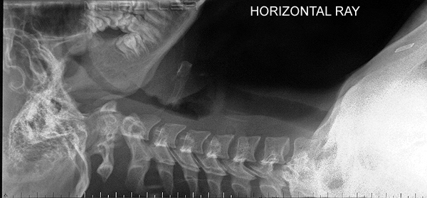
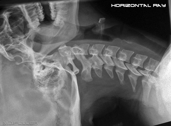
It is very easy to have the patient's mandible overly the upper cervical vertebra. Unfortunately, in a trauma situation, you may have little scope to change the position of the patient's mandible. The problem can be particularly evident in a patient who is in the wrong hard collar size/type, or if the hard collar is incorrectly fitted.
Shoulders
All of the upper endplate of T1 should be demonstrated on a lateral cervical spine image. The shoulders should be as inferior as possible. This can be very difficult to achieve in some patients. If the patient has a shoulder injury, the problem becomes worse. Having staff pull on the patient's arms/shoulders may help, but may also cause the patient to move out of a true lateral position, or may result in the patient being "pulled off the film/IR". The patient may also be injured by pulling on the patient's shoulders. Before pulling on the patient's arms, check with the supervising doctor. If it is the departmental policy to never pull on a patient's arms, your decision is already made.
In a flexible patient, particularly young females, you may be able to have the patient cross their arms across their chest. This will tend to position the humeral heads anteriorly allowing a less obscured demonstration of the cervico-thoracic junction.
SupervisingExposure TechniqueI find that the only way that I can be sure that....
- the patient's head is in the true AP position and.....
- the grid is vertical and.....
- there are no unnecessary artifacts and.....
- the shoulders are down as far as possible...
... is to don a lead gown, and stand at the patient's head end of the bed at the time of exposure. Refer to you department's policy on this one.
CollimationSome radiographers advocate a high kVp technique to maximise the chances of visualising the lower cervical vertebra. An aluminium wedge filter covering the mid and upper cervical vertebra can also be used to good effect.
RespirationThe collimation should include
- all of the cervical spine bony anatomy
- the soft tissues anterior to the cervical vertebra
- the cervico-thoracic junction
- the cervical spinous processes
- the clivus
The clivus is used to assess atlanto-axial alignment
SwallowingSome radiographers advocate exposure on expiration to maximise the inferior movement of the patient's shoulders. If the patient is ventilated, holding respiration during exposure will help to avoid movement unsharpness.
Soft Tissue VisualisationIt is helpful if the patient is not swallowing at the time of exposure. Swallowing can result in a false-positive retropharyngeal soft tissue swelling sign
If you are using non-digital (film-screen) equipment, it is important to utilise an exposure that demonstrates the bony and soft tissue anatomy. Important soft tissue signs of injury can be missed if the retropharyngeal soft tissues are overexposed.
Is it Lateral?
Critique
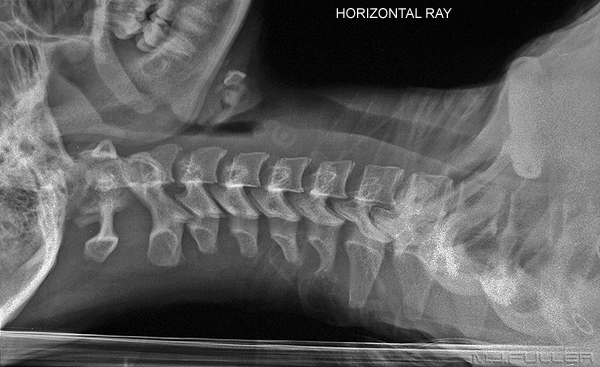
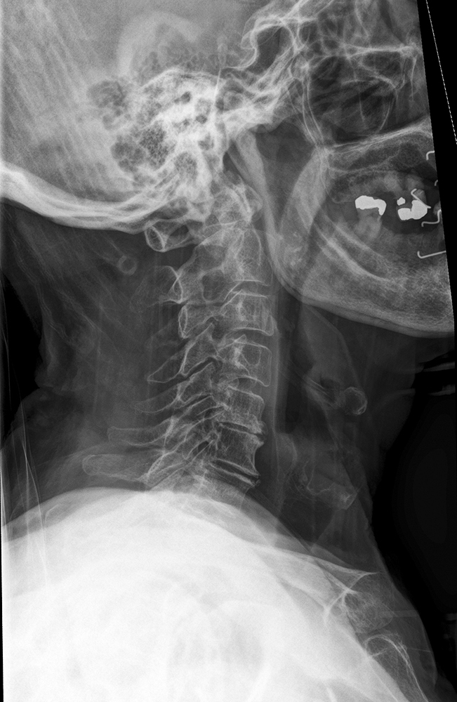
- The cervical spine is in a true lateral position
- The clivus is well demonstrated
- The upper endplate of T1 is not adequately demonstrated
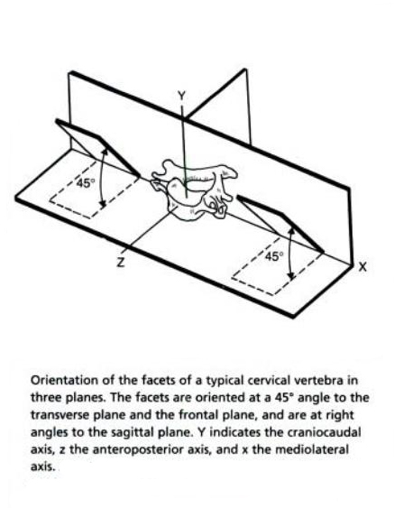
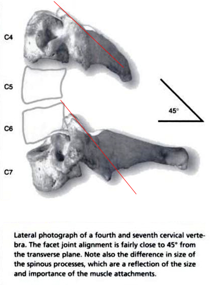
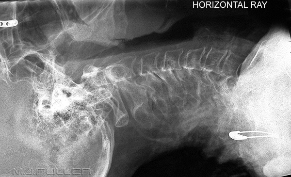
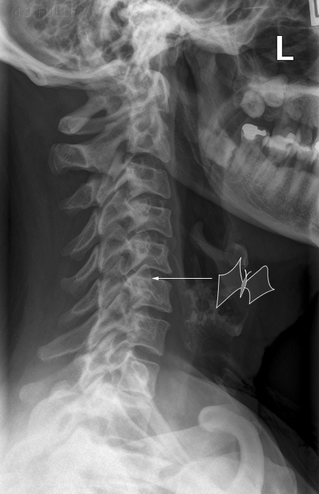
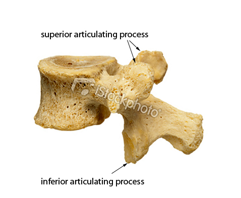
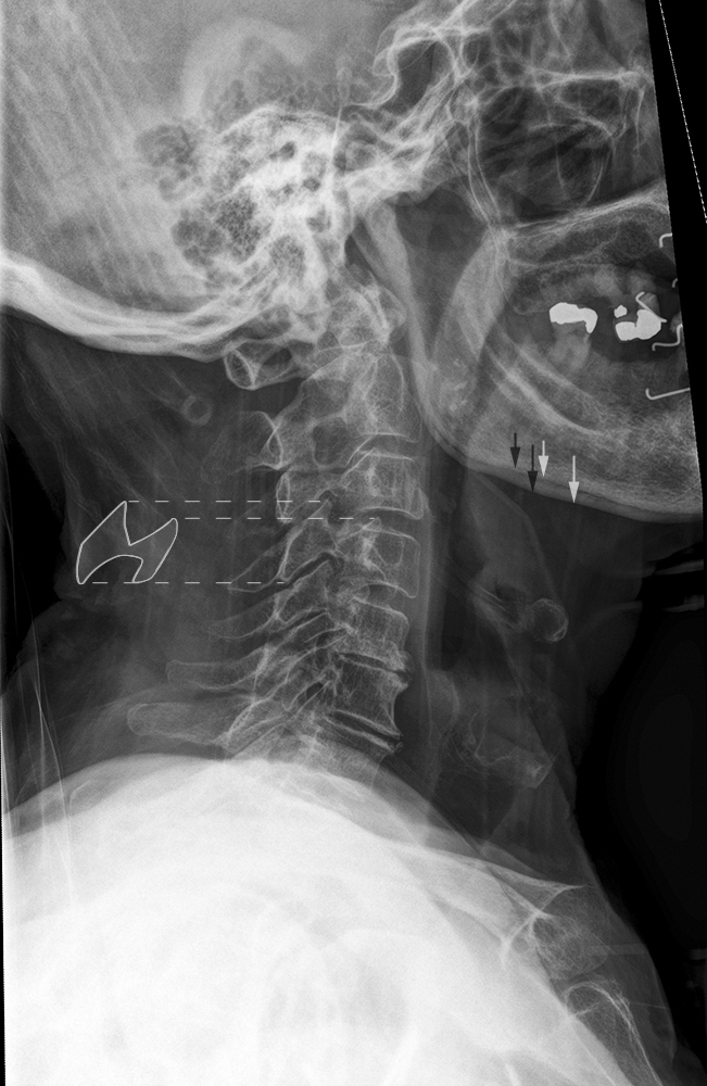
Producing a true lateral cervical spine image makes the job of image interpretation much easier. The butterfly sign is a reliable indication that the cervical spine is considerably malpositioned (off-lateral). The wings of the butterfly are the superior and inferior articulating processes.
It is noteworthy that despite the best attempts of the radiographer to position the patient in a true lateral position for cervical spine radiography, the resultant image will not always demonstrate a true lateral position. A failure to produce a true lateral cervical spine can be frustrating for the radiographer but is not necessarily blameworthy.
adapted from <a class="external" href="http://www.istockphoto.com/file_thumbview_approve/9979978/2/istockphoto_9979978-human-lumbar-vertebra-lateral-view.jpg" rel="nofollow" target="_blank">http://www.istockphoto.com/file_thumbview_approve/9979978/2/istockphoto_9979978-human-lumbar-vertebra-lateral-view.jpg</a>
....back to the applied radiography home page hereThe lateral cervical spine in the trauma room is still a required projection in many institutions. Compulsory routine CT scanning of the cervical spine in trauma patients will render this projection obsolete. Practise and diligence will yield results.
