The Skyline Patella Projection
There is nothing quite like nailing a perfect skyline knee projection. This page looks at some issues and tips for achieving the goal of perfect skyline knee radiography.
The Positioning Sponge
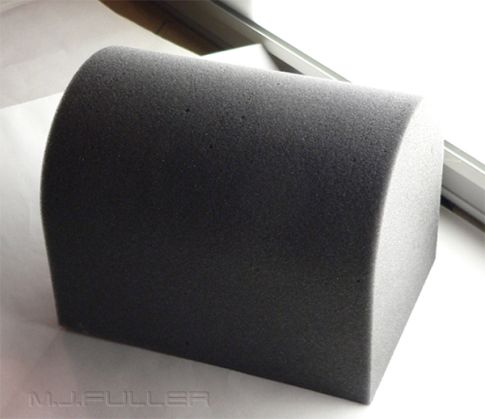
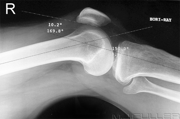
The "Research"In pursuit of the perfect skyline knee projection, I investigated the idea that the ideal tube angle might be more consistent if the knee flexion was always the same. It would also be desirable in a trauma situation if you could flex the knee slightly and rest it onto a positioning sponge such that it was positioned perfectly for a horizontal ray lateral using a 24 x 30cm (12 x 10 inch) cassette. Moreover, it would be desirable to leave the sponge in situ for the skyline view.
I had a large number of off-cut sponge pieces that I cut down to different heights until I achieved what appeared to be the perfect height. The final result is shown below.
Sponge can be cut with a hot wire or an electric carving knife. I made my own hot wire cutter. You can do a <a class="external" href="http://www.google.com/" rel="nofollow" target="_blank">google</a> search for more information if you want to make your own cutter. If you're not familiar with electrical safety issues, don't do it!
Alternatively, you can take your foam to a foam cutter for a professional looking result
The Image
Discussion
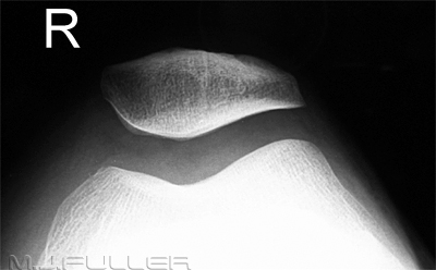
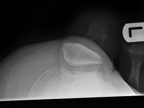
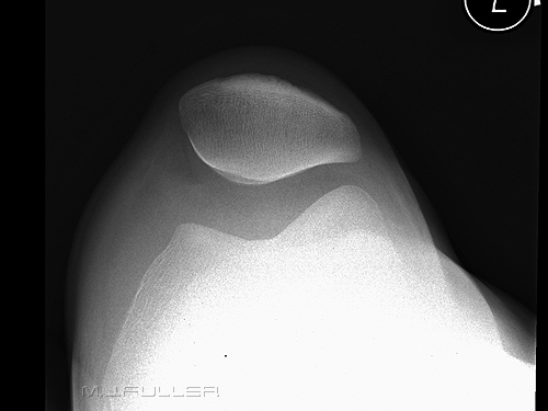
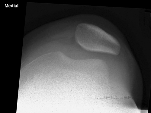
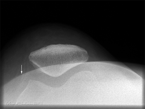
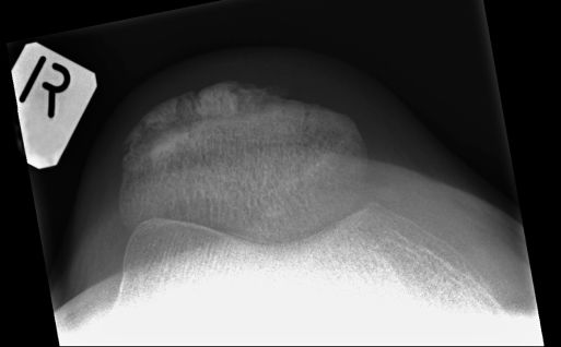
My understanding of the skyline patella position is that a minimum of knee flexion is more likely to reveal abnormal patellar tracking. The most frequent problem that I found with this technique was that there was too little knee flexion. Too little flexion of the knee joint can cause the tibial tuberosity or other anatomical structures to be projected over the anatomy of interest.
These images were produced with a CR system which allows for the easy measurement of angles. It would be possible to measure the skyline angle from the lateral view for all patients and you are likely to achieve very consistent quality images. The practicality of this approach is questionable.
Tips
The toes are in the way
It is good practice to remove the patient's footwear.
You have a number of choices if the patient's foot anatomy is superimposed over the patella.
- plantar flex the ankle joint like a ballerina "on point"
- increase knee joint flexion
- position the X-ray tube so that you are directing the X-ray beam along the medial or lateral side of the patient's foot
Radiographic TechniquesUnderexposureThe skyline projection requires considerably more radiation exposure than the AP/lateral. Also, the less flexion of the patient's knee, the more exposure you will need. Furthermore, for reasons of practicality, you could end up with a long FFD.
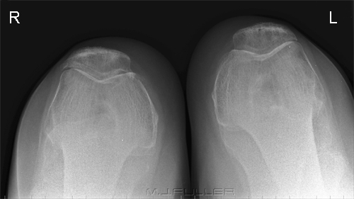
Comparison ViewsAs much as I dislike routine comparison views, there is justification for imaging both patellae in the one exposure. This is particularly useful when assessing subtle abnormal patellar tracking.
Supporting the cassette and patientSitting the patient up in a semi-sitting position is very uncomfortable. The DARRIN sponge is perfectly shaped to place behind the patient's back to provide support. One disadvantage of this approach is that the patient may receive a primary beam radiation dose to the orbits and thyroid.
My preferred technique is to keep the patient supine and use the LEDDRA skyline cassette holder.
Is the Skyline Projection Necessary?
What Went Wrong?
Case 1
Case 2
Case 3
Case 4
FaultCorrectionThis image is suffering form movement unsharpness.
This fault can be associated with technique. If you are using the "semi-sitting" patient position, it is best to put something behind the patient's back so they are not left "rocking" while they are supporting the cassette. I found that having the patient supine and using a cassette holder is a safer bet. See the page on the Leddra Skyline Cassette Holder.
Shorter exposure time will also help reduce movement unsharpness
Case 5
FaultThe soft tissue density (arrowed) that is arcing through the middle of the image is likely to be the soft tissues of the lower leg.
CorrectionIncrease knee flexion
Case 6
Case 7
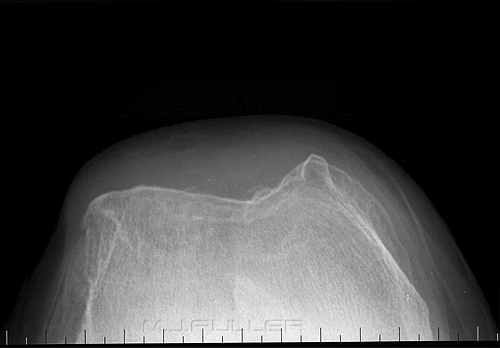
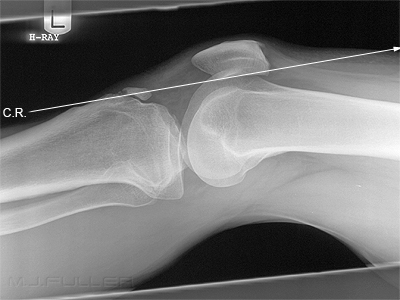
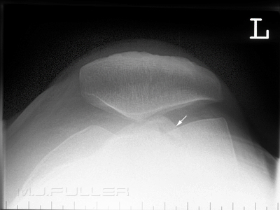
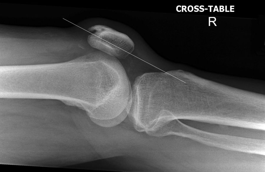
A skyline knee projection taken in this position, with the central ray as shown above, will project the tibial tuberosity over the patello-femoral joint.
Note- patient has Osgood-Schlatter disease .Resultant skyline patella image. The tibial tuberosity (arrowed) is projected over the patello-femoral joint. To correct this error further flex the patient's knee.
Case 8
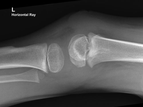
"The patella initially ossifies at between three and five years, commencing as multiple foci that rapidly coalesce" <a class="external" href="http://www.ncbi.nlm.nih.gov/pubmed/6729496" rel="nofollow" target="_blank">(Ogden JA. Skeletal Radiol. 1984;11(4):246-57. Radiology of postnatal skeletal development. X. Patella and tibial tuberosity.)</a>
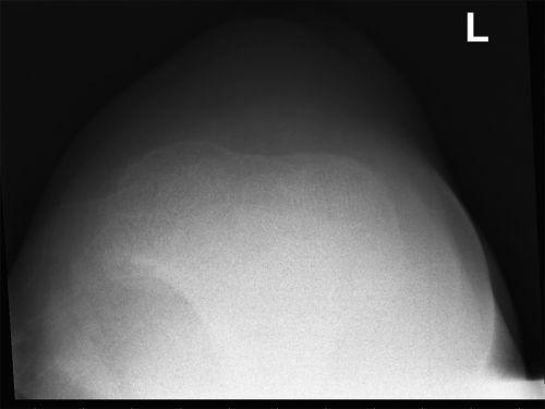
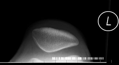
The skyline projection will not demonstrate a patella that has not ossified.Resultant skyline patella image.
Case 9
Case Studies
Case 1
Case 2
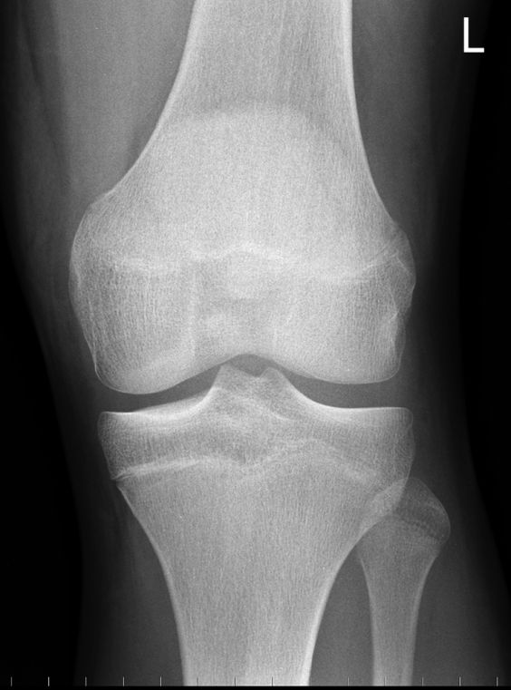
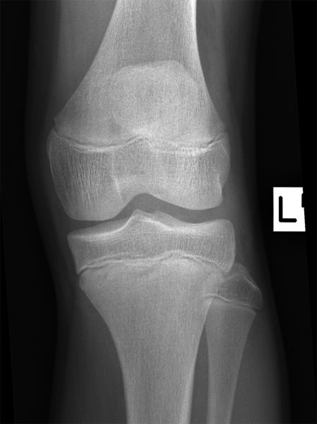
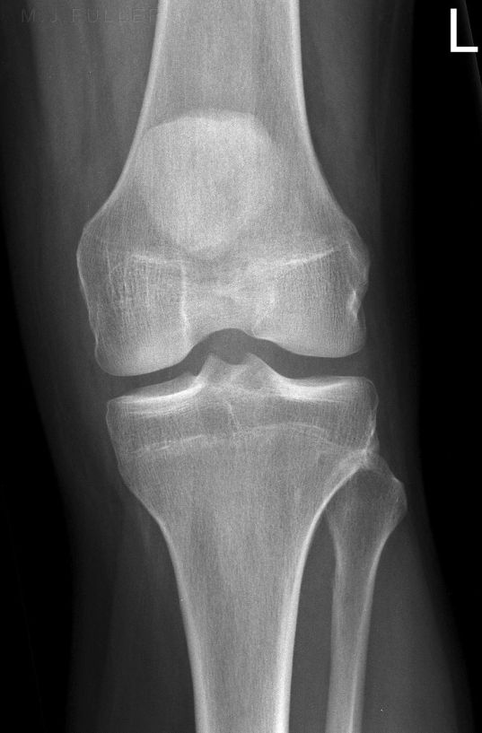
This 13 year old boy presented to the Emergency Department with a sore left knee following a sports injury. He was examined and found to be tender in the region of this left patellar tendon. He was referred for left knee radiography.
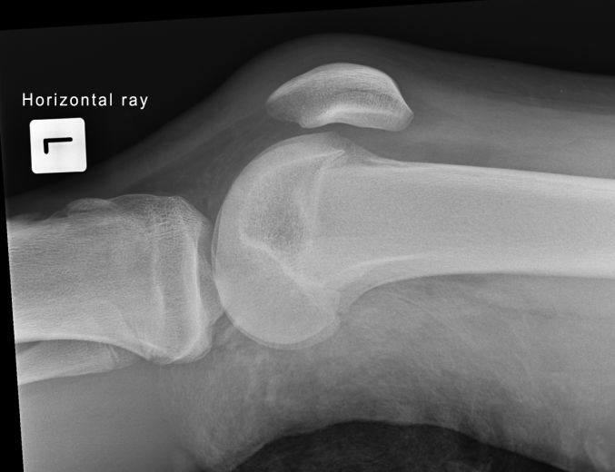
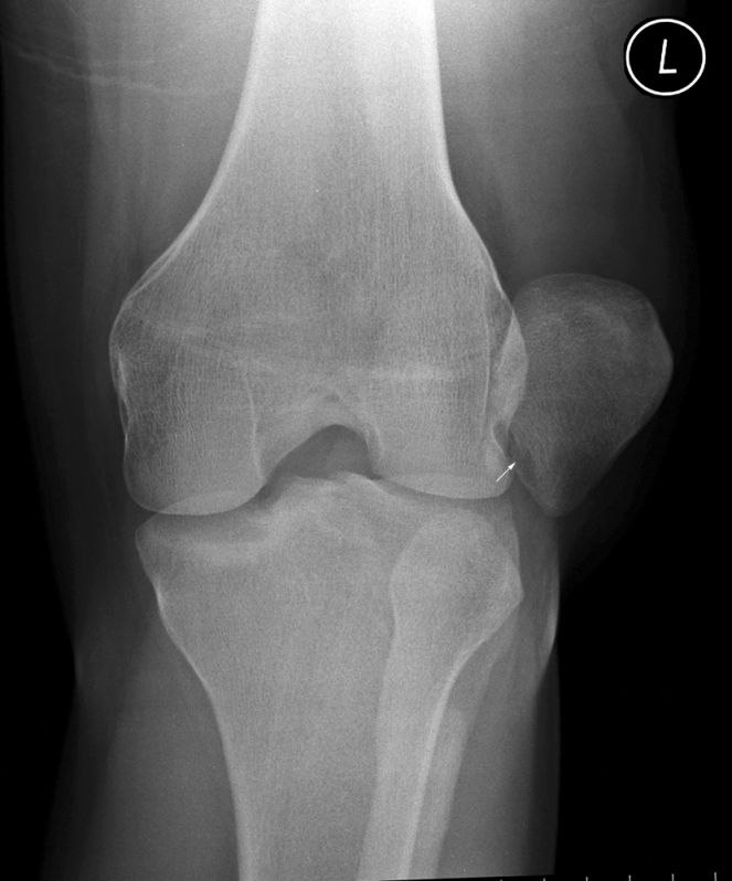
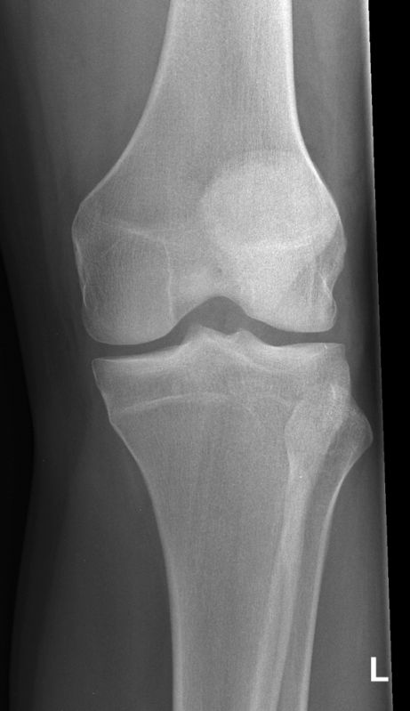
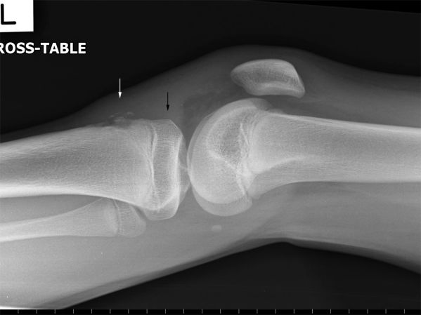
The AP knee image demonstrates no displaced fracture.The tibial tuberosity is fragmented (white arrow). Whilst this is not diagnostic for Osgood Schlatter's disease (OSD), the appearance is consistent with OSD.
Note the thickening of the patellar tendon with a possible fluid collection between the patellar tendon and the proximal tibial epiphysis (black arrow). Hoffer's fatpad demonstrates mixed fat and fluid density consistent with an acute injury.
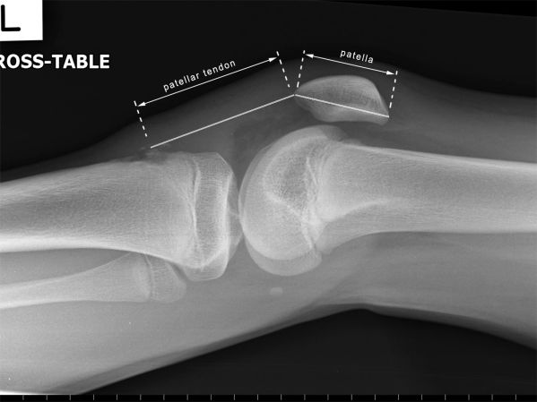
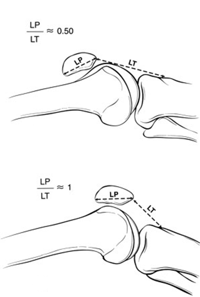
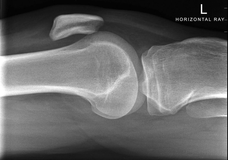
Patella alta notedThe position of the patella may be evaluated on the basis of the ratio of the greatest diagonal length of the patella to the length of the patellar tendon on lateral radiographs (the Insall-Salvati ratio). This measurement is relatively independent of knee flexion, and a ratio of less than 0.80 indicates patella alta
quoted from <a class="external" href="http://www.jbjs.org/Image_Quiz/2004/jun04/iqjun04_p2.shtml" rel="nofollow" target="_blank">The Journal of Bone and Joint Surgery
Image Quiz</a>
adapted from <a class="external" href="http://www.jbjs.org/Image_Quiz/2004/jun04/iqjun04_p2.shtml" rel="nofollow" target="_blank">The Journal of Bone and Joint Surgery
Image Quiz</a>The Insall-Salvati ratio in a normal knee (top) and in one with patella alta (bottom).
LP = length of the patella
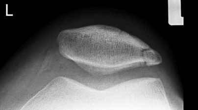
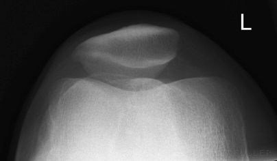
LT = length of the patellar tendon.The skyline knee projection demonstrates good patient positioning but the radiographer has had difficulty including all of the anatomy on the IR because of the patella alta.
Case 3
Case 4
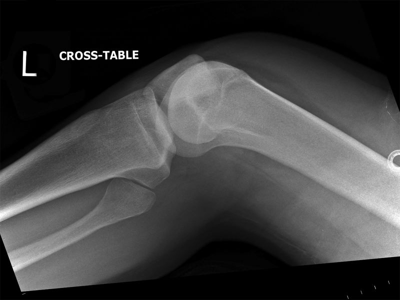
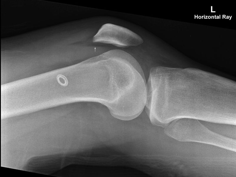
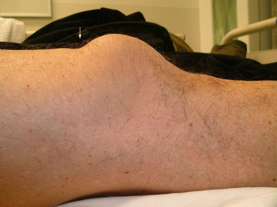
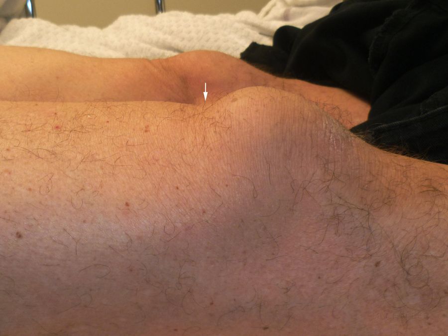
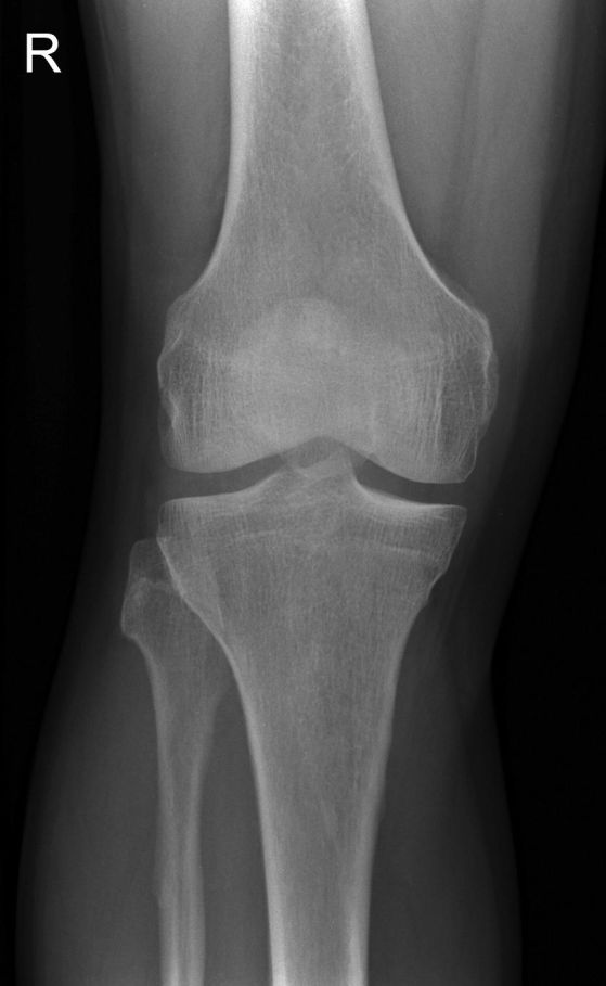
This 68 year old male presented to the Emergency Department following a fall onto a concrete floor. His right knee was painful and deformed with a very prominent patella. He described a hyperflexion injury to his right knee. He was referred for right knee radiography.
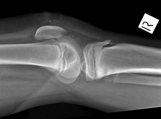
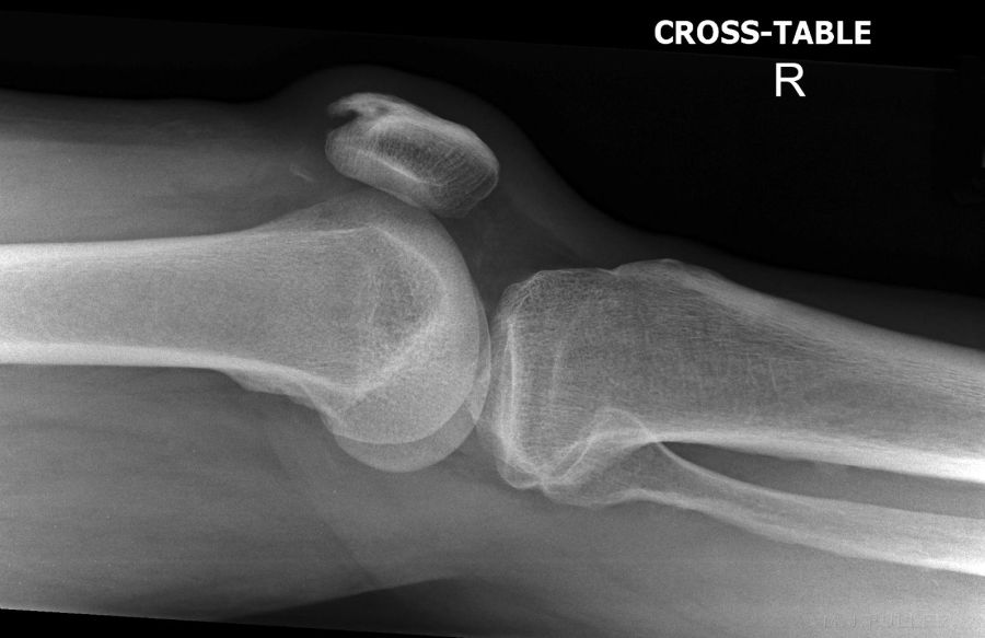
His right knee was seen to have a depression in the skin immediately superior to the upper pole of the patella. This was not evident on the contralateral side.The depression superior to the right patella(arrowed) was not evident on the contra-lateral side. The AP knee image is unremarkable. The lateral knee image demonstrates abnormal orientation of the patella. There is a some mixed density
within Hoffa's triangle. There is also loss of definition of soft tissue structures and mixed fat/fluid density in
the region of the suprapatellar pouch.
There is a well corticated bony density in the region of the suprapatellar pouch and a quadriceps tendon <a class="external" href="http://wiki.answers.com/Q/What_is_an_enthesophyte" rel="nofollow" target="_blank">enthesophyte</a>.The patella is pulled and tilted inferiorly by the action of patellar tendon in the absence of a countering
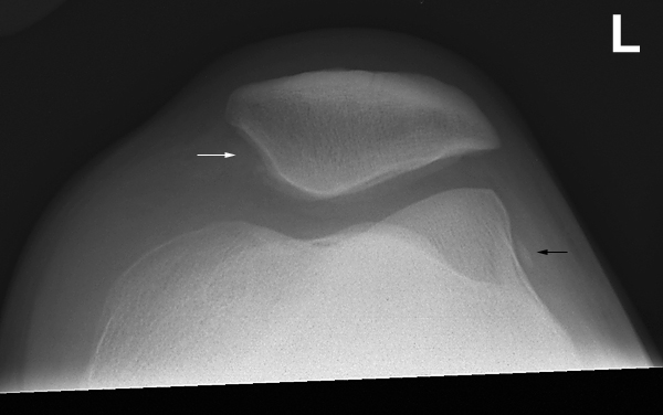
traction from the quadriceps muscle associated with a quadriceps tendon rupture.The skyline projection was destined for failure given the rupture of the quadriceps tendon
....back to the Applied Radiography home page here
