The Lateral Elbow

From wikiRadiography
Jump to navigation Jump to search
Introduction

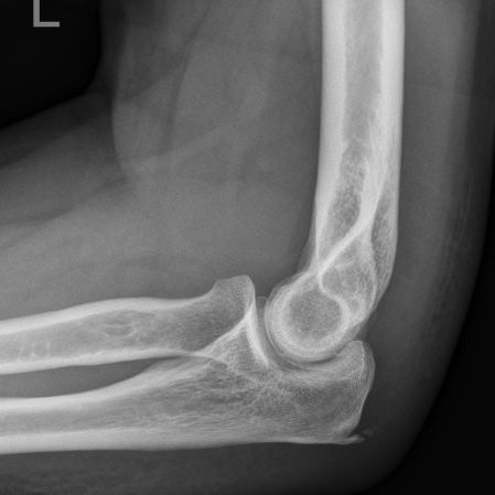
The lateral elbow is a troublesome radiographic position in terms of achieving a true lateral view. If you haven't achieved a true lateral view, understanding how to correct the position can also prove difficult. This page attempts to establish the anatomy that is displayed on the lateral view image and how to make position corrections when malposition occurs.

The Anatomy
Anterior Aspect of Distal Humerus
distal humerus
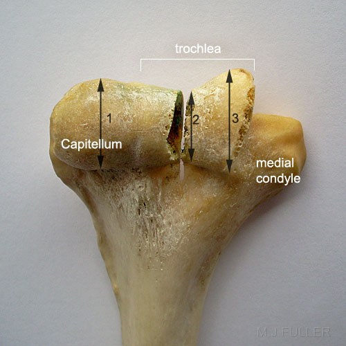
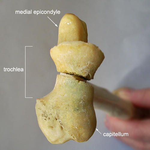
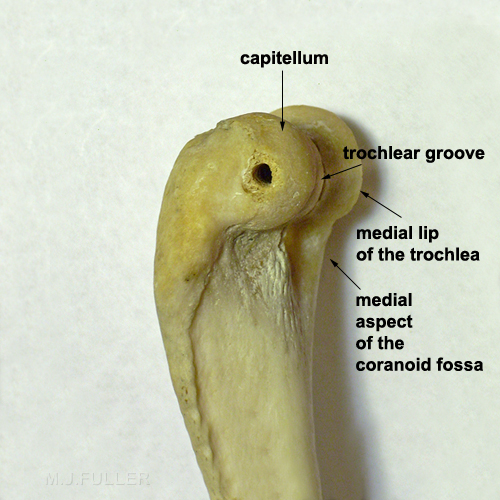
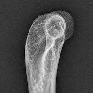
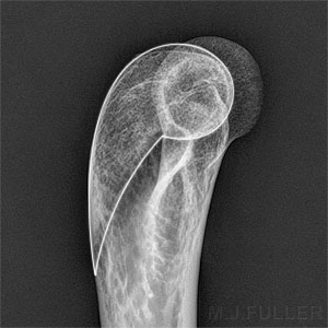
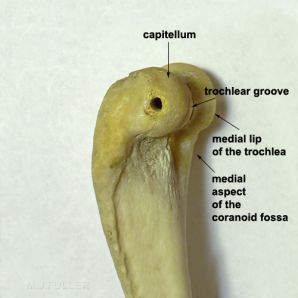
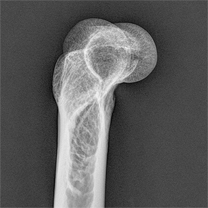
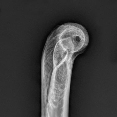
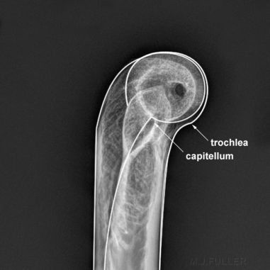
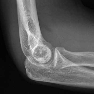
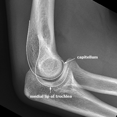
There are three circular bony structures that we aim to position concentrically when performing a lateral elbow radiographic position:

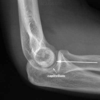
1. The Capitellum (approximately 19mm diameter)
2. The narrowest dimension of the trochlear groove (approximately 17mm diameter)
3. The widest dimension of the medial lip of the trochlea (approximately 22 mm diameter)

The diameter measurements above are were measured with a vernier caliper and rounded off to the nearest millimetre. They are the diameter measurement of the actual humerus photographed (left) and radiographed (below).


Note- this anatomical specimen has a saw cut through the trochlea (and a few rough edges)
Posterior Aspect of Distal Humerus
distal humerus
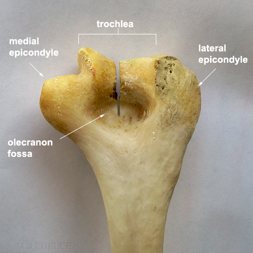
The olecranon fossa is deeper than the coranoid fossa.

The lateral condyle is larger than the medial condyle.


Note- this anatomical specimen has a saw cut through the trochlea
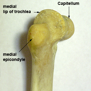
Medial Aspect of Distal Humerus
distal humerus
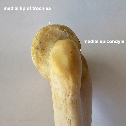
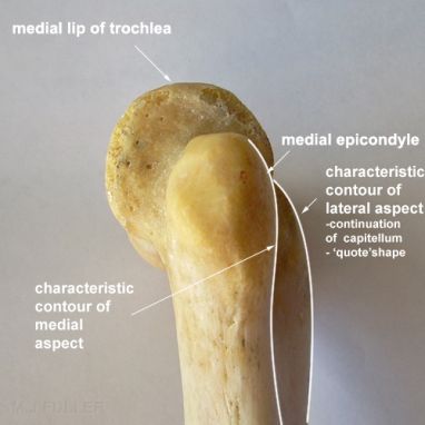
The medial aspect of the distal humerus shows the medial epicondyle positioned proximal to the trochlea.
Lateral Aspect of Distal Humerus
elbow anatomy


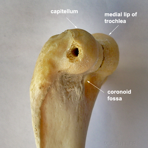
This anatomical specimen has a hole in the capitellum where it was attached to a complete skeleton.
Articular Aspect of Distal Humerus
distal humerus
This aspect of the distal humerus gives an impression of the larger lateral condyle compared to the medial condyle. You can also see that the capitellum articular surface in this view is a relatively short arc.


Note- this anatomical specimen has a saw cut through the trochlea


<embed allowfullscreen="true" height="350" src="http://wikifoundrytools.com/wiki/wikiradiography/widget/youtubevideo/723af899d4513a225691aff0242ff103110825d9" type="application/x-shockwave-flash" width="425" wmode="transparent"/>This is a video of the distal humeral X-ray anatomy in the lateral projection as the humerus is rotated through a limited arc from slightly externally rotated to slightly internally rotated.


Condyle, Epicondyle and Epicondylar Apophysis.

The distal humerus can be divided into two halves- the medial condyle and the lateral condyle in a manner analogous to the distal femur. "The articular portion of the medial condyle is the trochlea, and the articular portion of the lateral condyle is the capitellum. The epicondyle is considered part of the nonarticular portion of the condyle." <a class="external" href="http://emedicine.medscape.com/article/91780-overview" rel="nofollow" target="_blank">(Kelly, John D IV, e-medicine, Medial Condylar Fracture of the Elbow)</a> . The ossification centres of the medial and lateral epicondyles in a child are referred to as the unfused epicondylar apophysis.


Barium Coated Anatomy
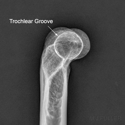
Trochlear Groove Distal Humerus
Trochlea
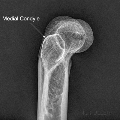
Medial Condyle Distal Humerus
medial condyle
The minimum diameter of the trochlear groove is relatively easy to identify on a lateral elbow because of its smaller diameter. The trochlear groove is outline with barium in this imageThe medial condyle is relatively easy to identify on the lateral projection given its characteristic shape and more proximal position relative to the capitellum and medial lip of the trochlea. The medial condyle is coated in barium in this image for ease of identification.


What is the Characteristic Humeral Appearances of a True Lateral Elbow?

lateral distal humerus
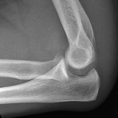
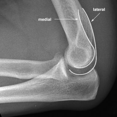
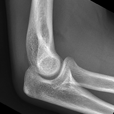
What is characteristic about the appearance of a true lateral elbow?
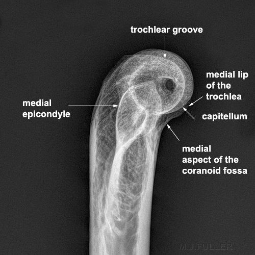
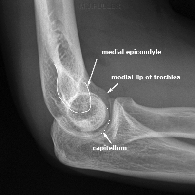
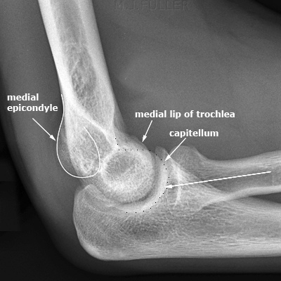
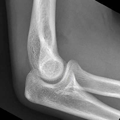
  • the trochlear groove cortex is concentric with, and of a smaller diameter than, the capitellum and medial lip of the trochlea- three concentric circles.
  • the medial epicondyle is slightly anterior rather than centrally located over the distal humerus
  • the figure 8 or hourglass shape is not characteristically positioned
  • the medial border of the distal humerus is approximately (but not perfectly) located in the middle of the distal humerus
  • the posterior continuation of the capitellum is the most posterior cortex demonstrated.
  • the appearance of the continuation of the medial lip of the trochlea (anteriorly) with the medial aspect of the coranoid fossa (as shown above) should not be considered a sign of malpositioning of the lateral elbow.
medial epicondylar poppy
adapted from <a class="external" href="http://www.moplants.com/gallery2/v/Her+Country+Garden_001/poppy.jpg.html" rel="nofollow" target="_blank">http://www.moplants.com/gallery2/v/Her+Country+Garden_001/poppy.jpg.html</a>
The medial epicondylar poppy!


The humerus poppy
lateral humerusThis photograph demonstrates the approximate position that the lateral view was taken in from a lateral perspective.


What are the Characteristic Humeral Appearances of a Malpositioned Lateral Elbow?
Too Externally Rotated
lateral elbow
Too Externally Rotated
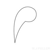
The Humerous Apostrophe
Bony Anatomy- lateral aspect
lateral humerus
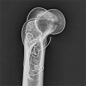
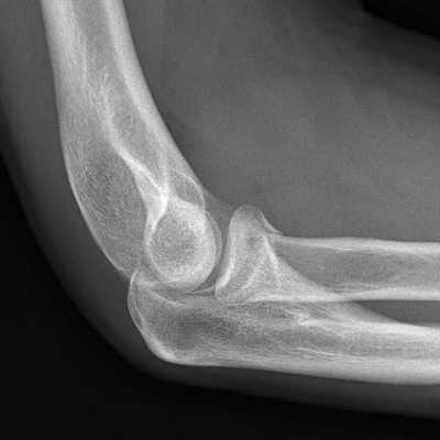
When the elbow is too externally rotated, the capitellum (side away from IR) rotates posteriorly. In doing so, the medial condyle moves anteriorly revealing an apostrophe shape of the capitellum and posterior cortex of the distal humerus.

To correct this malposition, lower the patient's hand.


Too Internally Rotated
distal humerus
Too Internally Rotated
The humerus elephant
adapted from <a class="external" href="http://www.math.utah.edu/%7Echerk/teach/elephant.GIF" rel="nofollow" target="_blank">http://www.math.utah.edu/~cherk/teach/elephant.GIF</a>
Bony Anatomy- medial aspect
the distal humerus
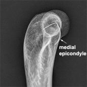
When the elbow is too internally rotated, the capitellum moves anteriorly and the medial condyle moves posteriorly. The appearance of the medial condyle as it appears posteriorly is a characteristic 'bump' in the posterior contour of the distal humerus. This is dumbo's head (medial epicondyle) starting to emerge.

To correct this malposition, raise the patent's hand off the IR

This was photographed from the medial aspect (opposite aspect to all of the other images) which may be a little confusing!


The Anterior Transition Points
lateral humeruslateral humeruslateral humerus

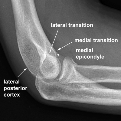
A subtle but useful method of differentiating the capitellum from the trochlea on the lateral view image is the difference in the shape of the transition points with the anterior aspect of the distal humeral metaphysis- the trochlea has a smoother transition than the capitellum. The trochlea transition is also visible on a true lateral elbow.

The Posterior Contours
lateral elbowlateral elbowlateral elbow
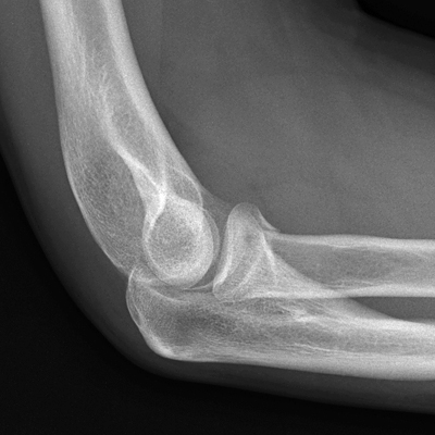
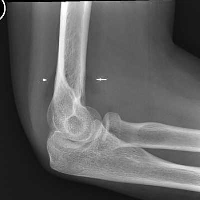
This is a malpositioned lateral elbow. The key to repositioning the elbow into a true lateral is in distinguishing the medial from lateral anatomy.The medial posterior cortical contour and the lateral posterior cortical contours are distinctly different.


The Proximal Radial Line as a Guide to Identifying the Capitellum.

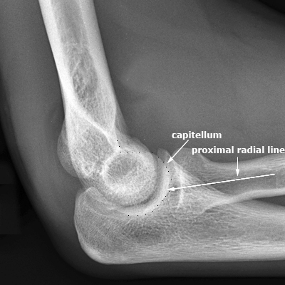
Up until this point, evaluation of the lateral elbow position has not included consideration of the proximal ulna, and more importantly, the proximal radius. Radiographers commonly use the proximal radial line to differentiate the capitellum from the medial lip of the trochlea on the lateral image. This is an important differentiation when attempting to correct a malpositioned lateral elbow.

lateral elbow
proximal radial line
If you were attempting to correct this lateral elbow position, the proximal radial line is a useful guide in differentiating the capitellum from the medial lip of the trochlea.

Note that this lateral elbow position is not necessarily worthy of a repeat view.
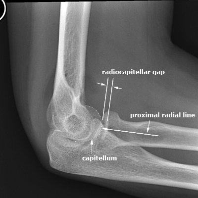
The proximal radial line is a line drawn through the middle of the radius that should bisect the capitellum. If it does not divide the capitellum into halves, it could indicate a dislocated radial head. Another useful feature of the proximal radial line is that it should always bisect the capitellum in any projection. The proximal radial line can therefore be utilised to identify the capitellum except when the radial head is dislocated. The proximal radial line essentially points to the capitellum.


The Radiocapitellar Gap as a Guide to Identifying the Capitellum

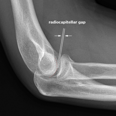
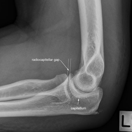
The radial head and the capitellum are covered in a layer of articular cartilage. These two articular cartilages tend to maintain a constant distance between the bony radial head articular margin and the bony capitellum. This radiocapitellar gap can be used as a guide to identification of the capitellum. This guide is particularly useful when the radial head is in true profile.

lateral elbow- radiocapitellar gap

lateral elbow- radiocapitellar gap
If you were attempting to correct this lateral elbow position, the radiocapitellar gap positively identifies the capitellum.The radial head is well profiled facilitating the capitellum using the radiocapitellar gap. The distal humeral structure overlying the radial head ifs the medial lip of the trochlea. The capitellum is separated from the radial head by a narrow gap- the radiocapitellar gap.


Testing The Theory

There will generally be two basic corrections for lateral elbow malposition

- raise hand/lower hand (external rotation/internal rotation of the humerus)
- raise/lower elbow (adduct/abduct humerus)


Case 1

lateral elbowlateral elbow- radiocapitellar gaplateral elbow
There is an anterior fatpad sign and a radial head fracture. The elbow is not in a true lateral position. The radiocapitellar gap identifies the capitellum as too posterior.


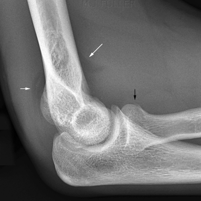
There is significant malpositioning. Whilst there are a number of indicators of the nature of the malpositioning, the 'humpback' appearance of the posterior cortex suggests that the patient's hand needs to be lowered.
lateral humerusThe Apostrophe
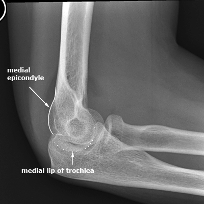
The medial epicondyle is projected too anteriorly

The 'humpback' appearance of the posterior cortex (quote sign) supports the suggestion that the lateral epicondyle is projected too posteriorly.

- lowering the patient's hand will result in the medial epicondyle moving posteriorly.

lateral elbow- lower hand


Case 2

lateral elbow
proximal radial line
lateral elbow
There is an anterior fatpad sign. The elbow is not in a true lateral position. The proximal radial line allows identification of the capitellum.The patient's hand position does not need to be corrected.

The capitellum is projected too distally. Raising the patient's elbow will correct this malposition.

lateral elbow- raise elbow


Case 3

LATERAL ELBOW
lateral elbow- compound errorlateral elbow- compound error
There are anterior and posterior fatpad signs. There is a radial head fracture. The elbow is not in a true lateral position. The proximal radial line allows identification of the capitellum. The radiocapitellar gap also supports the identification of the capitellum.The medial epicondyle is profiled posteriorly (dumbo's head sign)

This is a compound positioning error- the capitellum is projected
  • too distally (raise elbow to correct)
  • too anteriorly (raise hand to correct

lateral elbow- raise hand raise elbow


Case 4

lateral elbow
lateral elbowlateral elbow
There are anterior and posterior fatpad signs. The elbow is not in a true lateral position. The proximal radial line allows identification of the capitellum. The radiocapitellar gap also supports the identification of the capitellum.The medial epicondyle is profiled slightly posteriorly (dumbo's head sign)

This is a compound positioning error- the capitellum is projected
  • too proximally(lower elbow to correct)
  • too anteriorly (raise hand to correct

lower elbow raise hand


Case 5

lateral elbow
lateral elbowlateral elbow
There are anterior and possibly posterior fatpad signs. The elbow is not in a true lateral position. The elbow is not flexed to 90 degrees.The radiocapitellar gap identifies the capitellum (white dotted line) most convincingly. The medial epicondyle is not clearly demonstrated.The characteristic profiles of the medial and lateral condyles are outlined.

This is a compound positioning error- the capitellum is projected
  • too proximally(lower elbow to correct)
  • too posteriorly (lower hand to correct
lower hand, lower elbow


Case 6

LATERAL ELBOW
LATERAL ELBOWLATERAL ELBOW
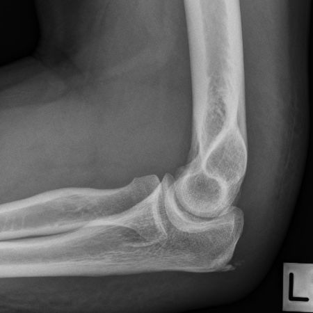
This male patient was referred for elbow radiography with an unexplained swollen and painful left elbow. The first attempt lateral elbow is shown above. The radiographer considered the elbow to be sufficiently malpositioned to warrant a repeat.The capitellum was identified using the radiocapitellar gap method. The radiographer used this method to establish that the patient's hand and elbow needed to be raised to achieve a true lateral elbow position.

This is a compound positioning error- the capitellum is projected
  • too distally (raise elbow to correct)
  • too anteriorly (raise hand to correct)
    lateral elbow- raise hand raise elbow
The results of the first (and only) attempt to correct the lateral malpositioned elbow are shown above. Ther three concentric rings are demonstrated indicating a true lateral elbow position


Comment

I was warned as a student that the lateral elbow position is the most difficult position in radiography. A knowledge of the elbow anatomy and the characteristic appearance of a malpositioned elbow will allow for a corrected position to be achieved. The usual cost-benefit balance should be considered before repeating a malpositioned lateral elbow view- just because you can achieve a corrected lateral elbow position doesn't mean that you should!

It is below a reasonable standard of care to repeat a malpositioned lateral elbow by randomly altering the elbow position and hoping for the best.


...back to the wikiradiography home page
...back to the Applied Radiography home page