The Lateral Ankle Trap
Jump to navigation
Jump to search
---- Case 1----Patient History
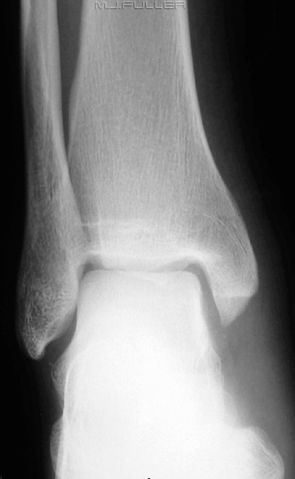
This patient presented to the Emergency Department with a painful and swollen ankle. The mechanism of injury was unknown. The referring doctor requested an ankle x-ray examination to rule out any bony injury.
Images
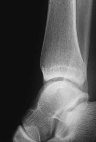
Image EvaluationThere is no apparent bony injury. An ankle effusion cannot be seen but the lateral ankle position and exposure do not afford a clear assessment. Is any further imaging warranted?
The radiographer has assessed the lateral ankle position as inadequate and proceeded to repeat this view.
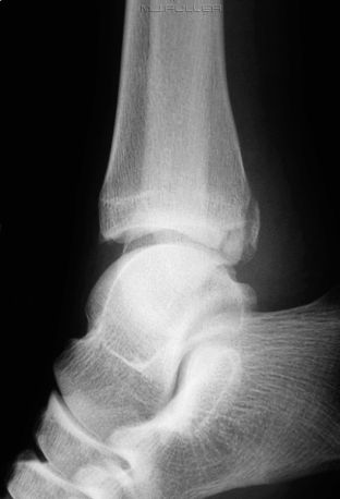
Repeat Lateral Ankle
The repeat lateral ankle has projected the distal fibula off the posterior malleolus of the distal tibia revealing a tibial posterior malleolus fracture.
DiscussionThe over-rolled ankle positioning error is particularly risky in terms of posterior malleolus fractures. It would be reasonable to ask if this is simply a freak occurrence?
---- Case 2 ----Patient History
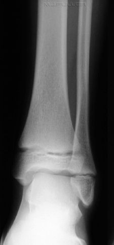
This child has presented to the Emergency Department following an unwitnessed fall. The patient is assessed and ankle X-ray imaging is requested
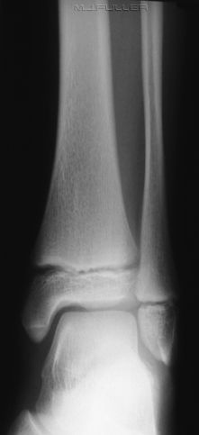
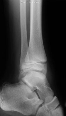
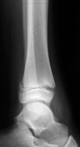
ImagesThe radiographer has performed an AP, Lateral and oblique X-ray examination and the images are shown below
Image EvaluationThe lateral ankle is over-rolled causing the distal fibula to be superimposed over the posterior malleolus. The radiographer considered this image worthy of repeating. The repeat image is shown below.
This lateral ankle is in a good position and reveals a Salter-Harris II fracture of the posterior distal tibia. (? SH I also)
DiscussionIt is worth considering the risks of obscuring a posterior malleolus fracture of the distal tibia in cases where the lateral ankle position is incorrect as shown in the two cases above. Where there is a high suspicion index of a fracture based on patient history, clinical presentation and soft tissue signs ( e.g. ankle effusion) a repeat view of the lateral ankle is highly recommended.
....back to the applied radiography home page here
