The Binocular Cone
Introduction
TerminologyAmong the many objectives that the radiographer must consider are scatter radiation reduction and patient dose reduction. The binocular cone aims to achieve both objectives in the one hit.
Light Beam Diaphragm.
The light beam diaphragm (LBD) is the apparatus attached to the X-ray tube housing that allows the radiographer to produce a variable sized parallel sided radiation field
Acromioclavicular Joint
The clavicle (collar bone) has two joints; the sternoclavicular joint medially and the acromioclaviular joint (AC joint) laterally
Polycarbonate
This is a type of strong clear thermosetting plastic sheet
Technique
ImagesIt is routine practice in many institutions to perform comparison views for acromio-clavicular joint injuries. There are two commonly performed techniques. The first technique involves a single AP slit exposure that includes both AC joints.
Technique 1
This method potentially produces very comparable images of the AC joints but irradiates the patient's thyroid unnecessarily
<a class="external" href="http://www.fadavis.com/related_resources/1_2037_693.pdf" rel="nofollow" target="_blank">http://www.fadavis.com/related_resources/1_2037_693.pdf</a>
Technique 2
The second technique involves imaging the normal and the comparison AC joint separately. This can be achieved using a single film by simply displacing the X-ray tube or displacing the cassette.
This tube-shift method does not unnecessarily irradiate the patient's thyroid, but, despite the radiographers careful positioning, can produce images that can lack comparability. This is probably attributable to the fact that the patients AC joints are never imaged in exactly the same projection/position.
Technique 3
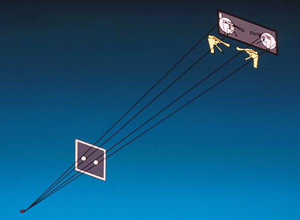
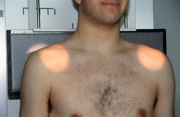
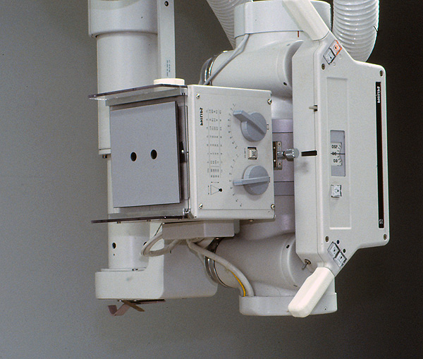
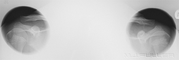
This technique uses a LBD mounted lead cone that produces two circular radiation beams. This method combines the benefits of the first two methods
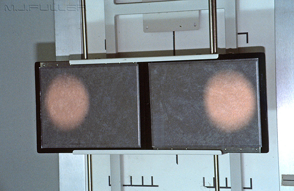
The technique is best achieved using a non-grid technique as shown below.
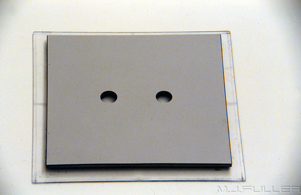
The cone is a simple device that is not that difficult to make. The device used here is a piece of 1mm lead sheet glued between two pieces of kitchen bench laminex. Two suitably sized and spaced holes are drilled in the laminated lead using a drill press. The holes are sized and spaced for an FFD of 180cm. The spacing and hole size can be determined using a mock up cardboard cone with two adjustable hole pieces.
It is best to mount the binocular cone on a piece of polycarbonate sheet that is custom cut to suit your light beam diaphragm. Hook and loop tape provides a simple mounting method. Experience has suggested that it is best to drill two matching oversize holes in the polycarbonate sheet so that the LBD light will not be attenuated by the polycarbonate. Mount the cone as shown below in the LBD.
If the patient's shoulders are too wide to fit both AC joints on a 35 x 43cm cassette, you can use two 18 x 24 cassettes as shown below.
It is good practice to fully utilize the LBD cones also. Once the binocular cone is mounted, using the LBD cones, cone in until you are sure that the adjustable LBD cones are almost matching the binocular cones. The reason for doing this is that some radiation will penetrate the binocular cone lead (albeit very small). In addition, this technique will tend to act like an extension cone because there will be semi-matching cones at several points along the X-ray beam.
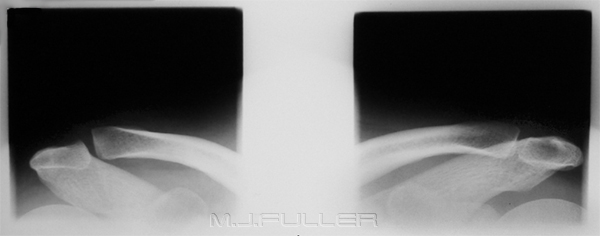
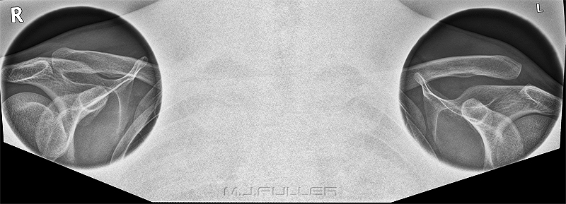
Alternate UsesYou should achieve an image that looks like the one below
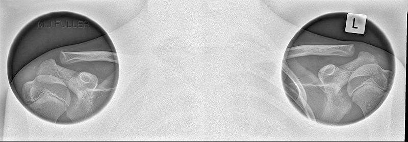
The binocular cone can be used on paediatric patients. Simply reduce the FFD and the radiation field become smaller and closer together. This paediatric patient had AC joint imaging using the same binocular cone as was used on the adult patient whose image is shown above.
TipsThe binocular cone does not have a wide variety of applications. One notable exception is shown below. These skyline knee images were taken on a morbidly obese patient who was referred for radiography of both knees. Whilst not ideally positioned, the images demonstrate that the binocular cone can be utilized to good effect for this projection.
It is important to include the coracoid in all AC joint imaging. An increased coraco-clavicular distance will indicate rupture of the coracoclavicular ligament
Even severe AC joint injuries can fail to be demonstrated on AC joint imaging. Take into consideration the mechanism of injury and the appearance of the shoulder profile. If an AC joint injury is clinically suspected and not revealed, repeat the examination weight-bearing or in an AP sitting position with the patient's hands placed across opposite knees.
Discussion
The binocular cone has been in use in my department for approximately 10 years. It is popular amongst the radiographers for its ease of use and effectiveness as well as aesthetic considerations. As with all LBD mounted devices, care should be taken to ensure that it does not fall onto the patient. It is good practice to lock it in place, or attach a tether cord.
Remove the cone from the LBD when the examination is completed.
....back to the applied radiography home page here
