The Abdominal Plain Film- Intramural Gas
Jump to navigation
Jump to search
Introduction
Aetiology
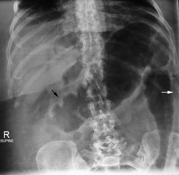
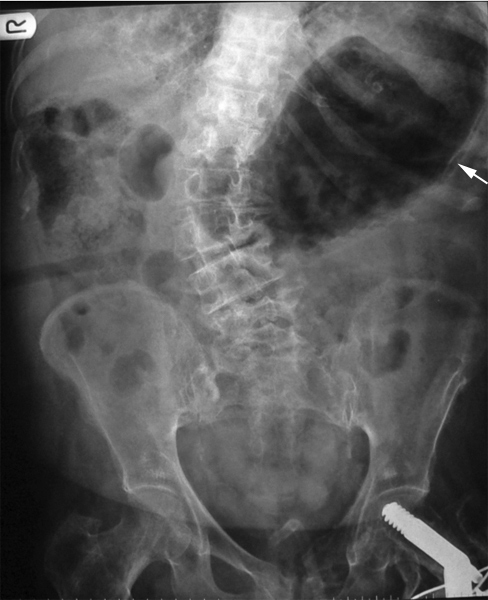
Toxic Megacolon
Case 1
Case 2
Case 3
Case 5
Conclusion
....back to the Applied Radiography home page
Introduction
Intra-mural gas refers to abnormal gas in the wall of hollow abdominal viscus. Intra-mural gas is one of the most serious findings on abdominal plain film requiring timely surgical intervention in adults. Intra-mural gas is not a difficult pattern recognition challenge. Radiographers should be able to identify mural gas to facilitate timely treatment.
Aetiology
Linear streaks of intra-mural gas indicate infarction of the bowel wall. Gas in the wall of the bowel in the neonatal period, whatever its shape, is diagnostic of necrotising enterocolitis. (<a class="external" href="http://www.medstudents.com.br/radio/radio2.htm" rel="nofollow" target="_blank">http://www.medstudents.com.br/radio/radio2.htm</a>)
Toxic Megacolon
Toxic megacolon should be considered one of those life-threatening conditions that can be diagnosed on plain abdominal film. If untreated, the mortality rate is between 20 and 30%. (Stephen R. Baker, The Abdominal Plain Film, Appleton and Lange, 1990 p203) Toxic megacolon is a fulminant form of colitis. Plain film appearance is of moderately dilated bowel with abnormal haustral pattern. The moderately dilated bowel will not dilate to the extent seen in sigmoid volvulus. There is usually no faeces in the bowel. The presence of intra-mural gas can quickly advance to perforation. These changes are most often demonstrated in the transverse colon.
Ulcerative colitis is the most common cause of toxic megacolon.
The characteristic features of toxic megacolon are acute dilation of a segment of colon, florid inflammation of the wall of the colon. The colon wall may be thickened in areas marked inflammation or thin and fragile in areas of colon where the mucosa has broken down and sloughed off. The colon wall in affected area may lose its characteristic pattern of haustra and plicae. The wall may be thickened and pseudopolyps may protrude into the lumen.
The transverse colon was thought to be a common site of toxic megacolon. This may not be the case- it may be that the transverse colon is where the disease is visualised on plain film because of the presence of gas (the transverse colon is anterior and tends to fill with gas when the patient is in the supine position). This would suggest that a decubitus projection would be advantageous to demonstrate the full extent of the diseased colon- left and right decubitus abdomen projections may also be warranted.
Barium enema is contra-indicated in fulminant colitis rendering the plain film an even more important diagnostic tool.
Case 1
Case 2
Case 3
Case 4
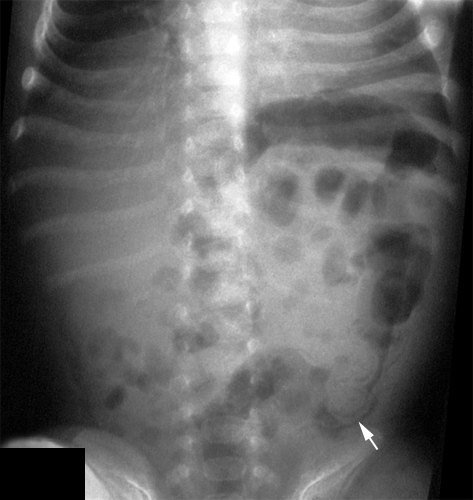
<a class="external" href="http://myweb.lsbu.ac.uk/dirt/museum/margaret/758-267c-1080241.jpg" rel="nofollow" target="_blank">http://myweb.lsbu.ac.uk/dirt/museum/margaret/758-267c-1080241.jpg</a>"Clinical presentation:
Male 3 weeks old, blood and mucus in stool. There is gas distension of transverse colon and splenic flexure, proximal to an area of abnormal bowel in the descending colon. There is the irregular double line of gas in the bowel wall (white arrow), separate from bowel contents. Immediately above the left iliac crest, there are two biconcave triangles of gas density. These extend above to a line of gas that is (left) lateral and separate from the line of intramural gas. Gas is seen on both sides of the abnormal colonic wall. The anterior location and the AP. projection mean that the transverse colon can be projected above the more posterior fundus of the stomach."
<a class="external" href="http://myweb.lsbu.ac.uk/dirt/museum/p7-abdo.html" rel="nofollow" target="_blank">myweb.lsbu.ac.uk/dirt/museum/p7-abdo.html</a>
Case 5
<a class="external" href="http://myweb.lsbu.ac.uk/dirt/museum/margaret/758-267c-1080241.jpg" rel="nofollow" target="_blank">
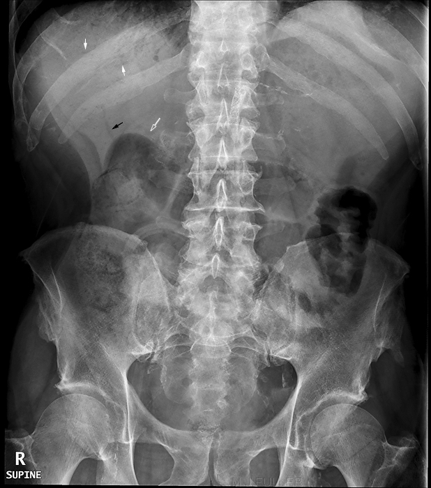
</a>"Clinical presentation: 67 year old male with renal failure and previous ileostomy. Recent SMA stenting. ? SBO
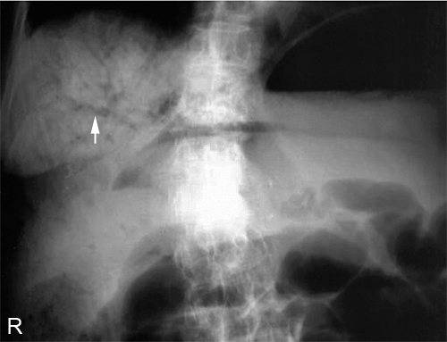
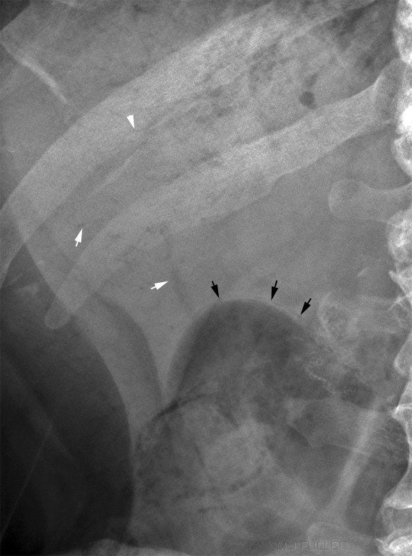
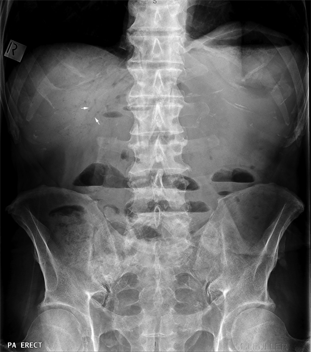
There is evidence of hepatic-portal venous gas (solid white arrows). There are dilated loops of small bowel. There is a prominant loop of gas-filled small bowel on the left with evidence of wall thickening. The black arrow identifies what is probably fat within the gall-bladder bed. The white outline arrow identifies what may be portal venous gas.This is a magnified view of the supine abdominal plain film. In addition to the hepatic-portal venous gas (white arrows) there is evidence of intramural gas (black arrows). The lucent structure identified by the bottom white arrow is probably fat around the gall bladder. The erect abdominal plain film demonstrates extensive hepatic-portal venous gas (white arrows). There are multiple airfluid levels within the bowel suggesting a motility disorder. <img align="bottom" alt="portal venous gas" src="http://image.wikifoundry.com/image/1/jGWwjd2Ap2NdY6Fd58WtVA240521" title="portal venous gas"/> This is a magnified view of the erect abdominal plain film RUQ. The hepatic portal venous gas is clearly seen as well as intramural gas (black arrow) and surgical sutures (white arrow).
Conclusion
Intra-mural gas is one of the more serious findings on abdominal plain film. A timely report by the radiographer to the referring doctor of the possibility of intra-mural gas could improve the prognosis for the patient. Decubitus views should be considered where there is a possibility of more extensive disease and where the additional information will affect patient management.
....back to the Applied Radiography home page
