The Abdominal Plain Film- Gasless vs Featureless
Introduction
Gasless and featureless abdominal plain films have a 'look of the abnormal'. This page examines the difference between gasless and featureless patterns and their significance.
Definition
A gasless abdominal plain film refers to an absence or minimum of gas in the gastrointestinal tract.
A featureless abdominal plain film is one in which there is little or no visualisation of the normal abdominal viscera.
The Gasless Abdominal Plain Film
Gas in the gastrointestinal tract can commonly accumulate from two sources. Firstly, gas in the stomach and small bowel can be ingested with food. Some patients habitually air swallow or may air-swallow when in pain. Gas in the large bowel can be endogenous, resulting from fermentation processes of faecal material.
An absence of gastrointestinal gas on abdominal plain film is not specifically abnormal (but is suspect). However, consideration should be given to the possibility of a gasless obstruction. Equally, a check of the patient history may reveal relevant information such as total colectomy.
Causes•A gasless abdomen could indicate•Patient is not an ‘air-swallower’•Mesenteric ischaemia•Obstruction of the stomach or oesophagus•Persistent vomiting from conditions such as pancreatitis or gastroenteritis
Normal Gasless Small Bowel
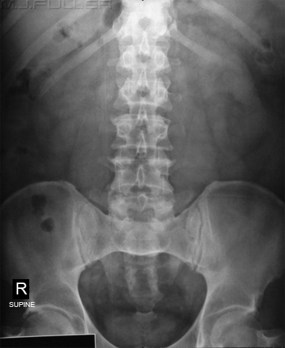
This patient appears to have a gasless small bowel. Fluid-filled loops of small bowel are present but are never as well visualised as air-filled loops.
There is debate regarding what is a normal amount of small bowel gas. Gas enters the small bowel during eating and drinking. Some people habitually air-swallow while others air-swallow at times of stress or when they are in pain. If the patient is air-swallowing at a higher rate than the small bowel can absorb the air, it will be visualised in the bowel. Some texts will claim that up to seven fluid levels in the small bowel on an erect abdominal film can be normal. Most texts quote a more conservative figure (2 is common).
Don't confuse gasless with featureless. A featureless abdomen can be a result of tumour or ascites. This patient has a gasless rather than featureless abdomen. Note that the renal, liver, psoas muscles and urinary bladder outlines are visualised
Note also that this abdominal film is not guaranteed to be normal. It could represent an early gasless small bowel obstruction. Clinical correlation is required.

Gasless Small Bowel Obstruction
You could be forgiven for thinking that this patient has been drinking dilute gastrografin. This appearance is a gasless small bowel obstruction and the opaque looking small bowel loops (white arrow) are filled with normal succus entericus, and/or ingested fluid, rather than gastrografin. If you compare this image with the gasless small bowel image above you can see that this small bowel is significantly more prominent.
There can be difficulty in distinguishing an early gasless small bowel obstruction from a normal appearance of the small bowel in someone who has just eaten a large meal.
Clinical correlation and follow-up imaging will usually provide confidence in the diagnosis.
The large bowel is not clearly visualised suggesting that it may be collapsed.

Colectomy
The Featureless Abdominal Plain Film
This patient has Crohn's disease and has had a previous colectomy. Note the absence of the large bowel. Note also a faint suggestion of a stoma in the left iliac fossa.

Ascites
This patient has prominent loops of gas-filled small and large bowel. The normal abdominal viscera are not demonstrated. Apart from the prominent bowel, the abdomen is featureless. The cause of this appearance is a large quantity of ascites. Note that the liver, spleen, kidneys, psoas and urinary bladder outlines are not seen. The reason that the bowel is somewhat centrally located is that it is floating in the ascites. If you are suspicious that the patient has air-filled bowel floating in ascites, it can be useful to perform an erect or decubitus view. This left lateral decubitus view demonstrates that the air-filled bowel floats up to the right flank which is the least dependant part of the abdominal cavity in the left lateral decubitus position.
Note that this is not a "right decubitus"- The 'right' annotation refers to the right side of the abdomen and the "decubitus" annotation refers to the patient position.
Tumour


Mass Effect
The CT abdomen shows the mass (arrowed) sited anterior to the psoas muscles.


Blood- AAA
Summary
The aneurysmal abdominal aorta is visible (arrow) because of its calcified wall. The abdominal viscera are not well visualised. The psoas, renal and kidney outlines are not well demonstrated. This raises the question of whether the AAA is leaking blood into the peritoneal cavity.
Blood- Ruptured Viscus
The supine abdominal plain film demonstrates a featureless pattern. There are no clearly defined psoas, kidneys or spleen. A CT abdominal scan revealed extensive laceration of his left kidney and spleen.
Artifact is external to patient


Gasless and featureless plain abdominal films often strike the observer as having an unusual appearance. It is useful to be able to distinguish between those images that are likely to be normal from those that are not. As with all abdominal plain film imaging, the appearance of the abdomen on plain film can have greater meaning when placed in the context of patient history, clinical presentation and other test results.
back to the applied radiography home page