Supine Intercondylar Knee Radiography
| Introduction |  |
At some stage in your career as a radiographer you will be asked to perform an intercondylar knee view on a patient who cannot easily and/or safely adopt the kneeling position. For some radiographers this is a daily event!
The kneeling intercondylar view of the knee joint can be self-defeating in that the patients who need it the most are least likely to be able to adopt the kneeling position. One of the solutions to this problem is to perform a modified AP intercondylar view by partially flexing the knee and utilizing one or more positioning sponges to locate the X-ray cassette. Four enterprising radiography students (Susie Curran, Steve Parker, Cameron Moore and Melita Greaney) from the University of Queensland in Australia decided that there was probably a better way and set about designing a positioning aid for AP supine intercondylar knee radiography. The result was simply brilliant!
The DARRIN
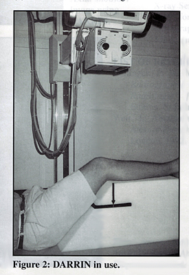
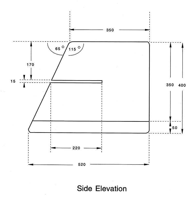
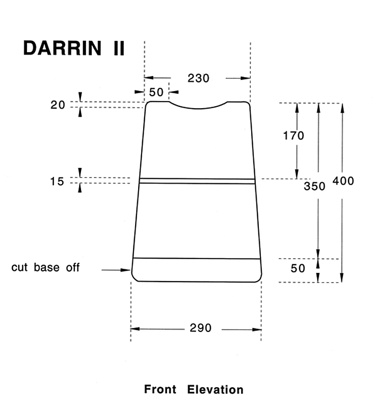
| DARRIN is an acronym for Device for Accurate Reproducible Radiography of the Intercondylar Notch The designers published their research results in The Radiographer (vol. 44: No. 1, April 1997 pp37-39). The DARRIN is pictured in figure 2 as it appeared in that original article. Using the DARRIN the patient's knee tends to fall into the correct position every time it is used. It is largely foolproof- once you have used it a few times you will become accustomed to positioning the patient such that the intercondylar notch is projected into the middle of the cassette. My experience over many years has been that the DARRIN lives up to its name- the results are accurate and reproducible. The DARRIN was not a commercial product at the time that it was published in The Radiographer so I set about having some made (with the consent of the designers). The plans I used are shown below. These are my original drawings that I submitted to the foam cutting company. The foam I used was a grey 29/400 (this refers to the density of the foam). If your foam converter doesn't have a computer controlled cutter, it would be best to seek one out for this job. |
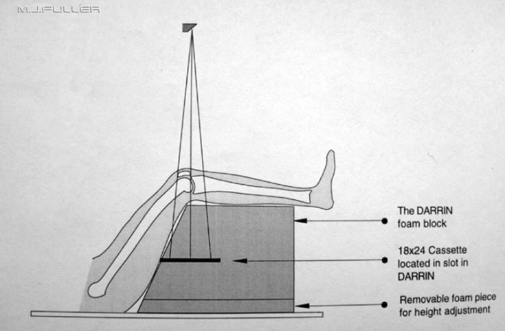
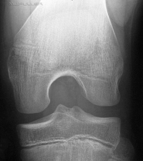
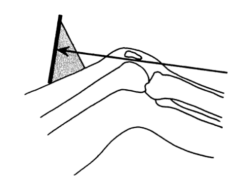
The patient is positioned supine on the X-ray table. An 18x24 X-ray cassette is placed transversely in the slot in the DARRIN. The patient's hip and knee are flexed and placed resting on the DARRIN as shown below. Centre the X-ray beam to the inferior border of the patella.
Tips
- Take care to ensure that the patient's knee is not rotated
- For tall patients, you may need to add the removable foam piece (see diagram above)
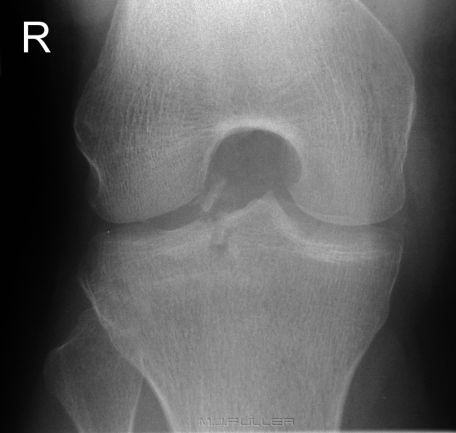
| | One of the unexpected features of intercondylar knee radiography with the DARRIN was the apparent comfort afforded by the device reported by patients with significant knee injuries. This image was achieved with the aid of the DARRIN. At the time the image was taken, it was not known that the patient had sustained an avulsion fracture of one of the tibial spines. The patient found the position so comfortable that he did not want to remove his leg from the DARRIN |
Images
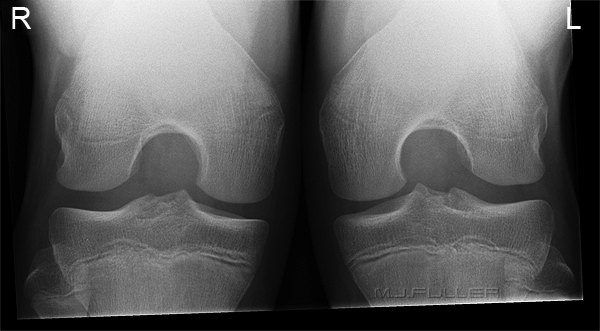
If you follow these instructions you should end up with an image as shown belowThis particular DARRIN design is for single knee radiography using an 18 x 24cm (10 x 8 inch) cassette. The image below demonstrates a bilateral intercondylar knees image using the DARRIN with a 24 x 30cm (12 x 10 inch) cassette.
Advantages/Disadvantages of the DARRIN
By any fair assessment, the advantages of the DARRIN far outweigh the disadvantages
Advantages
- easy to use
- consistent results
- suitable for injured patients
- improved patient safety
- can be utilised for other radiographic positioning
- inexpensive
Disadvantages
- there is a theoretical increase in geometric unsharpness associated with the increase in object-film distance (OFD). This is not detectable on the images
- there is a theoretical risk of cross-infection if blood enters the open cell foam. In practice this has not caused a problem- the DARRIN is covered in HDPE plastic film if there is a risk of blood staining or simply not used with these patients
- There is a need to increase the X-ray exposure to account for the increased in OFD. This increase in dose is partly offset by a very low repeat rate and should be balanced against the improved result
Other Applications
If you give a Radiographer a new positioning sponge, there's a fair chance he/she will find some new uses for it. Amongst the alternate uses for the DARRIN that radiographers have developed are the following
- If the DARRIN is turned up-side-down, it can used to support the contra-lateral leg in horizontal ray lateral hip radiography
- the DARRIN can be used for the SI shoulder projection
- The DARRIN can be used to produce a modified SI shoulder view in injured patients. Simply lean the patient back on the DARRIN and use a vertical central ray
- The DARRIN can be used for patients to lean on when performing skyline knee radiography
Modified SI Shoulder with the DARRIN
The superior-inferior (SI) shoulder is an important view of the shoulder. Unfortunately, as occurs commonly in radiography, it can be self-defeating; when you need it the most the patient is least likely to be able to adopt the correct position.
This patient presented with an anterior dislocation of the gleno-humeral joint. The dislocation was reduced and the Radiographer performed AP and lateral scapula views to demonstrate the effectiveness of the relocation. The orthopaedic surgeon was not entirely satisfied that the relocation had been confirmed and requested an SI projection. In these cases, the SI projection can be achieved with as little as 15 degrees of abduction of the humerus. The image may not be pretty but it does demonstrate anatomical alignment. In this case, the radiographer was faced with the prospect of achieving an SI projection of the shoulder in a patient with who was not prepared to contemplate any abduction of his humerus. This is where the radiographer's metal is tested in the Emergency Department; there is often a work-around, you just have to find it.
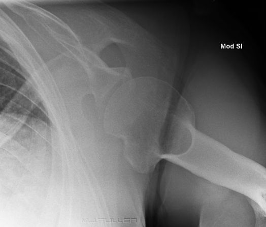
The radiographer decided to use the DARRIN to produce a modified SI projection. The patient simply leaned back on the DARRIN and a vertical X-ray beam was employed. The result is shown below
This projection is approximately 20 degrees off a true SI shoulder. The orthopaedic surgeon was very satisfied with the image.
It is noteworthy that the DARRIN can be used for conventional SI shoulder radiography as well as this modified technique.
Hill-Sachs lesion noted.
Skyline Knees Patient Back Support
The skyline view of the patella can be performed by employing a variety of techniques. One common technique is to position the patient supine and rest the cassette on the patient's thighs. In order for the patient to hold the cassette in this position, they either need long arms or they need to adopt a half-sitting position. This is very uncomfortable an almost impossible to hold for any length of time. The DARRIN can be employed to good effect in supporting the patient's back.
Alternatively, the patient can lie supine and support the cassette with a Leddra Skyline Cassette holder.
....back to the applied radiography home page here
...back to the Applied Radiography page
