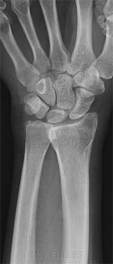
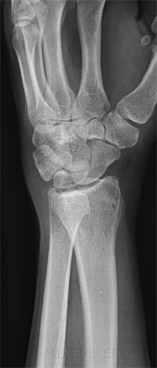
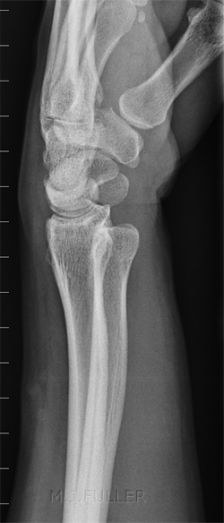
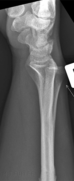
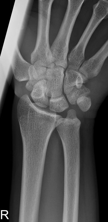
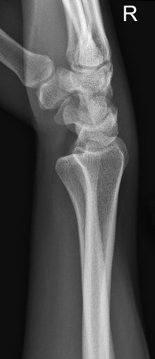
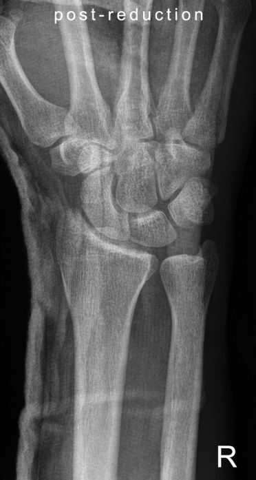
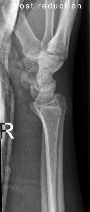
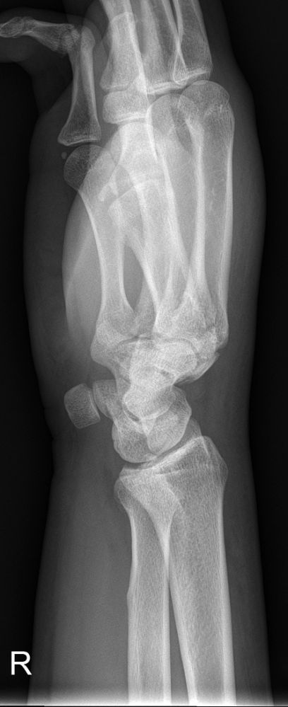
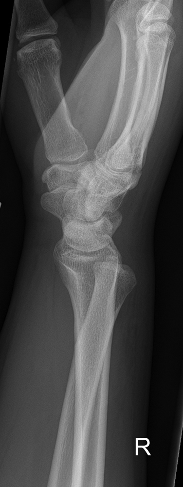
Subluxation of the distal radioulnar joint
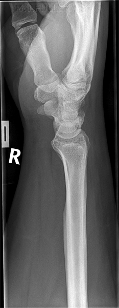
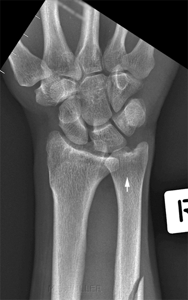
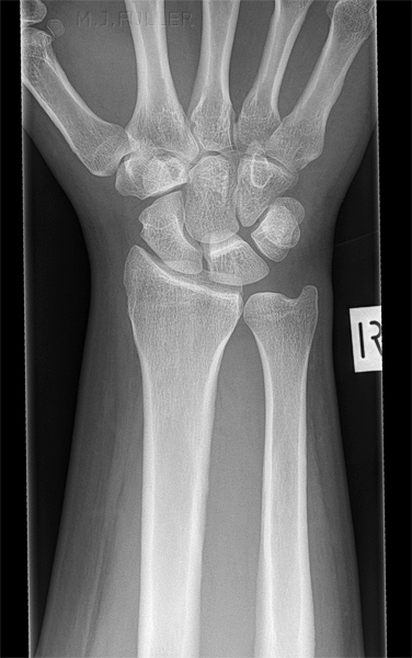
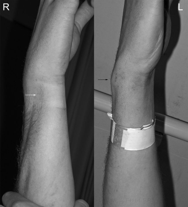
Dislocation of the distal radioulnar joint (DRUJ) without concomitant fracture is an uncommon injury. DRUJ subluxation is very easy to miss in the first presentation. It would appear that a common cause of misdiagnosis is to dismiss the appearance as positional/projectional.
Clinical Presentation
<embed height="350" src="http://widget.wetpaintserv.us/wiki/wikiradiography/widget/youtubevideo/4a8ad50f420d4685d6b17b53aae089afe4794af1" type="application/x-shockwave-flash" width="425" wmode="transparent"/>
Anatomy
"Distal ulna has a convex articular surface; this articulates with the concave semicylindrical sigmoid notch of radius. The important stabilizers of the distal radio-ulnar joint include all of the separate structures composing the triangular fibrocartilage complex. Of great clinical importance is the fact that these structures blend at the fovea, thus creating the potential for instability of the distal radio-ulnar joint when the ulnar styloid process is fractured. The flat pronator quadratus muscle originates from a long, narrow strip of the volar aspect of the distal part of the ulna and has a broad insertion on the volar aspect of the radius. It acts as a secondary stabilizer of the DRUJ by providing compressive force across the joint during pronation and supination."
<a class="external" href="http://www.rcsed.ac.uk/fellows/lvanrensburg/classification/wrist/distalradioulnajoint.htm" rel="nofollow" target="_blank">http://www.rcsed.ac.uk/fellows/lvanrensburg/classification/wrist/distalradioulnajoint.htm</a>
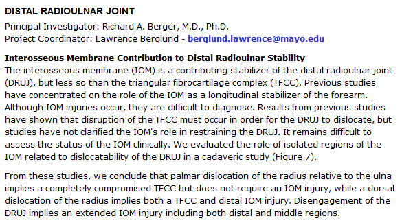
A study of the role of the interosseous membrane on the stability of the DRUJ reported the following
Radiographic Features of DRUJ Subluxation
- widening of RU joint on AP view;
- fracture (or non union) at base of ulnar styloid;
- significant shortening of the radius;
- obvious dislocation on the lateral view;
- it is essential that the lateral view be taken w/ proper technique so that the radial styloid process overlies the proximal pole of the scaphoid, lunate, and triquetrum;
- when proper positioning is ensured, dorsal or volar subluxation is noted by the relative position of the ulna above or below the radius;
<a class="external" href="http://www.wheelessonline.com/ortho/radial_ulnar_joint_instability" rel="nofollow" target="_blank">http://www.wheelessonline.com/ortho/radial_ulnar_joint_instability</a>
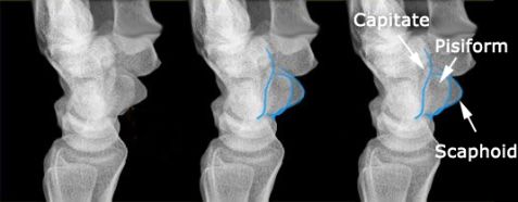
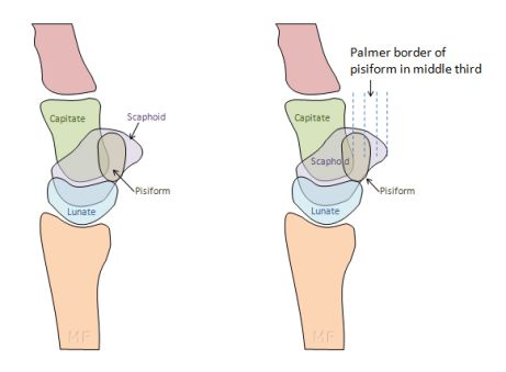
What is a Lateral wrist?
adapted from <a class="external" href="http://www.radiologyassistant.nl/en/476a23436683b" rel="nofollow" target="_blank">http:/</a><a class="external" href="http://www.radiologyassistant.nl/en/476a23436683b" rel="nofollow" target="_blank">/www.radiologyassistant.nl/en/476a23436683b</a>
quoted from <a class="external" href="http://www.radiologyassistant.nl/en/476a23436683b" rel="nofollow" target="_blank">http://www.radiol</a><a class="external" href="http://www.radiologyassistant.nl/en/476a23436683b" rel="nofollow" target="_blank">ogyassistant.nl/en/476a23436683b</a>
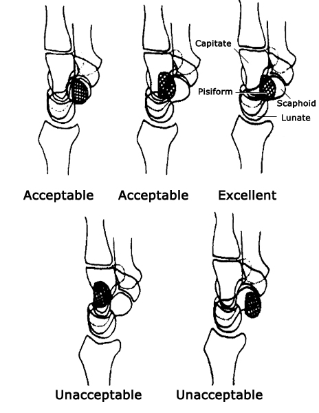
drawing based on
<a class="external" href="http://radiology.rsnajnls.org/cgi/content/full/219/1/11" rel="nofollow" target="_blank">Charles A. Goldfarb, MD, Yuming Yin, MD, Louis A. Gilula, MD, Andrew J. Fisher, MD and Martin I. Boyer, MD
</a><a class="external" href="http://radiology.rsnajnls.org/cgi/content/full/219/1/11" rel="nofollow" target="_blank">What the Clinician Wants to Know</a>
<a class="external" href="http://radiology.rsnajnls.org/cgi/content/full/219/1/11" rel="nofollow" target="_blank"> Wrist Fractures: What the Clinician Wants to Know1 </a>
<a class="external" href="http://radiology.rsnajnls.org/cgi/content/full/219/1/11" rel="nofollow" target="_blank"> Radiology. 2001;219:11-28.</a>
adapted from
<a class="external" href="http://radiology.rsnajnls.org/cgi/content/full/219/1/11" rel="nofollow" target="_blank">Charles A. Goldfarb, MD, Yuming Yin, MD, Louis A. Gilula, MD, Andrew J. Fisher, MD and Martin I. Boyer, MD
</a><a class="external" href="http://radiology.rsnajnls.org/cgi/content/full/219/1/11" rel="nofollow" target="_blank">What the Clinician Wants to Know</a>
<a class="external" href="http://radiology.rsnajnls.org/cgi/content/full/219/1/11" rel="nofollow" target="_blank"> Wrist Fractures: What the Clinician Wants to Know1 </a>
<a class="external" href="http://radiology.rsnajnls.org/cgi/content/full/219/1/11" rel="nofollow" target="_blank"> Radiology. 2001;219:11-28.</a>These authors go one step further by establishing the bounds of acceptability of lateral wrist positioning.
Case 1
This 45 year old lady presented to the Emergency Department after falling onto her wrist.
Case 2
Case 3This 38 year old male presented to the Emergency Department with a right forearm injury after a steel pipe fell on his arm.
Case 4
Discussion
Isolated subluxation/dislocation of the DRUJ is an easily missed diagnosis. The positive ulnar variance was a firm and consistent sign of wrist injury in the 2 cases. If the radiographer has identified the possibility of a DRUJ injury, appropriate supplementary views, including a PA and/or lateral view of the contra-lateral wrist may be useful.
...back to the Applied Radiography home page
