Soft Tissue Signs- The Ankle

From wikiRadiography
Jump to navigation Jump to search
Introduction

An equivocal fracture appearance should be considered in the context of other information such as

  • Patient history
  • Clinical presentation
  • Radiographic soft tissue signs


This page will examine the radiographic soft tissue signs of bony injury of the ankle- what they are, their plain film appearances, their limitations, and their utility.


Ankle Effusion- The Teardrop Sign

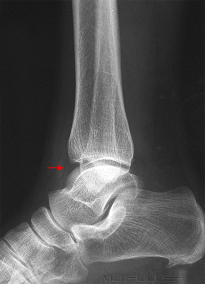
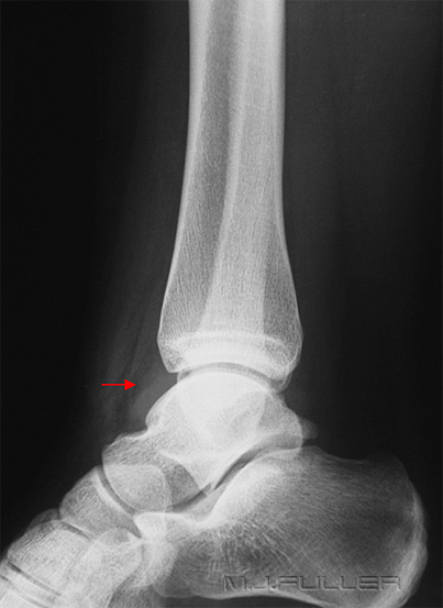
An ankle effusion suggests a significant injury to the ankle joint. The anterior and posterior extra-capsular region of a normal ankle joint should appear as a fat-like density. In the presence of an ankle effusion, the capsule can become distended and may appear to have a more fluid-like density


Soft Tissue Signs in Orthopaedics - wikiRadiography

Soft Tissue Signs in Orthopaedics - wikiRadiography
normal ankle demonstrating a fat -like density (arrowed) abnormal ankle demonstrating a fluid-like density (arrowed)


ANKLE EFFUSIONankle effusion
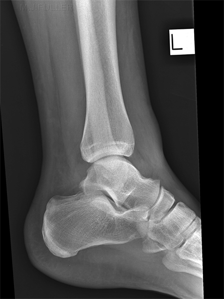
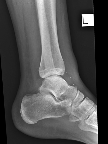
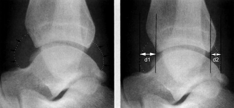
This patient presented to the ED following fall. There is extensive soft tissue swelling. There is an ankle effusion with the anterior and posterior recesses visibly distended with fluid. Same image with the anterior and posterior ankle joint recesses marked. Note that it is difficult to be sure of the exact contour of the posterior ankle joint recess. In the absence of an ankle joint effusion, the area bounded by the dotted line would show a fat density rather than a fluid density


Soft Tissue Signs in Orthopaedics - wikiRadiography
<a class="external" href="http://www.ajronline.org/cgi/reprint/134/5/985.pdf" rel="nofollow" target="_blank">Richard Towbin1, J. Scott Dunbar1, Jeffrey Towbin2, Robert Clark3. Teardrop Sign: Plain Film Recognition of Ankle Effusion. AJR:134, May 1980</a>

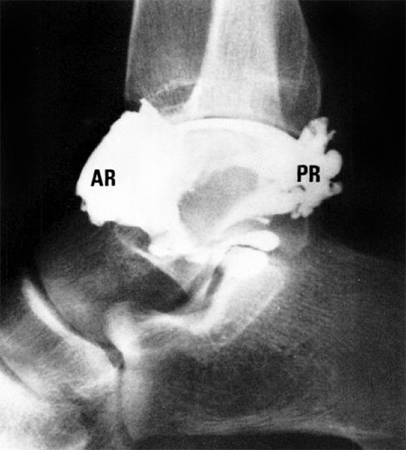
This is an ankle arthrogram which simulates an ankle effusion. The distension of the anterior recess (AR) and posterior recess (PR) of the ankle joint are simulated.

"(ankle) Fat pads are consistently visualized on the plain radiograph. Anteriorly the pretalar fat pad usually appears crescentic, and is cradled in the neck of the talus. There are two posterior fat pads, a thin juxtaarticular and a larger pre-
Achilles fat pad. The juxtaarticular fat pad is closely apposed to the posterior recess. It appears as a thin, linear structure about 1 -2 mm wide, that courses in a nearly straight line from the posterior aspect of the tibia to the posterior superior part of the calcaneus. The larger posterior fat pad is the triangular, pre-Achilles fat pad" (1)


Ankle Effusion- Correlation with occult ankle fracture

Clark et al (1) investigated the correlation between an ankle effusion and occult ankle fracture. They concluded that the presence of an ankle effusion suggested underlying fracture. Furthermore, they quantified the correlation as follows.


Soft Tissue Signs in Orthopaedics - wikiRadiography
adapted from Timothy W. I.Clark1, Dennis L. Janzen, Kendall Ho2, Anton Gnunfeld2, Douglas G. Conneli’
Detection of Radiographically Occult Ankle Fractures Following Acute Trauma : Positive Predictive Value of an Ankle Effusion AJR 1995;164:1185-1189
Carke et al (2) reported that "An ankle effusion of I 3 mm (d1 + d2) or greater in total capsular distension has a positive predictive value of 82% for occult fracture and is a reasonable threshold to prompt additional imaging."

References

1. Richard Towbin1, J. Scott Dunbar1, Jeffrey Towbin2, Robert Clark3. Teardrop Sign: Plain Film Recognition of Ankle Effusion. AJR:134, May 1980

2. Timothy W. I.Clark, Dennis L. Janzen, Kendall Ho, Anton Gnunfeld, Douglas G. Conneli’. Detection of Radiographically Occult Ankle Fractures Following Acute Trauma : Positive Predictive Value of an Ankle Effusion AJR 1995;164:1185-1189


Extra-articular soft tissue Swelling
ankleankle
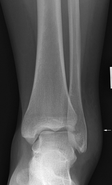
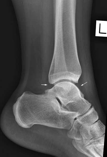
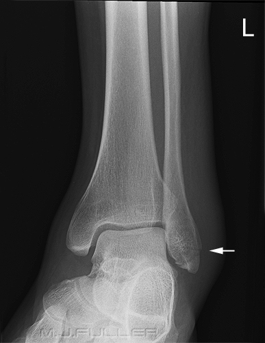
This 18year old female presented to the ED following a sports injury in which she "rolled her ankle". The AP ankle view image demonstrates extensive soft tissue swelling over the lateral malleolus. The lateral ankle image shows soft tissue swelling around the ankle joint. The anterior and posterior recesses of the ankle joint maintain a fat-like density suggesting that there is no ankle joint effusion.


Soft Tissue Swelling over Malleoli

NORMAL ANKLE SOFT TISSUES

ankle soft tissue swelling
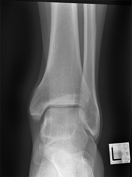
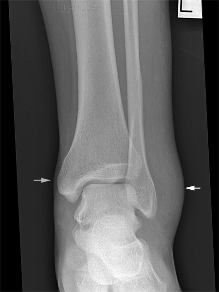
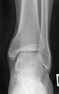
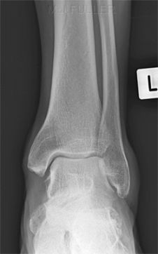
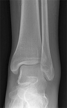
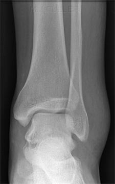
This is what you would expect to see in a normal ankle- the soft tissue overlying the medial and lateral malleoli are similar in thickness. It's not the thickness of soft tissue overlying the malleolus that counts per se, it's the symmetricality. i.e. an obese patient may have very a great thickness of soft tissue over the malleoli, but if it is the same on both sides, it would be considered normal.This patient presented to the ED after twisting his ankle. Note the extensive soft tissue swelling over the lateral malleolus- compare with the normal soft tissue thickness medially


Ankle APankle soft tissue
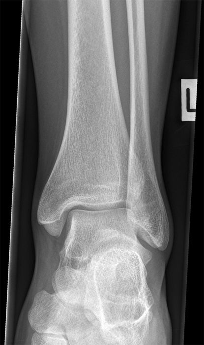
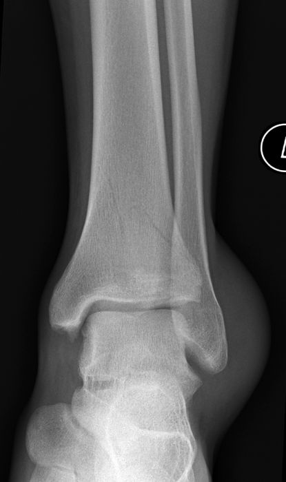
Normal symmetrical ankle soft tissuesThis patient has soft tissue swelling over the lateral malleolus. This is likely to be associated with a ligamentous injury- the bony injury is to the distal tibial metaphysis.


Soft Tissue Swelling over the Lateral Malleolus

Soft tissue swelling over the lateral malleolus is relatively common because inversion injuries of the ankle are relatively common. An extreme amount of soft tissue swelling does not necessarily indicate a fracture is present. On the contrary, it is frequently seen in severe sprain injuries (tendon/ligament injuries). In equivocal cases where you are suspecting a lateral malleolus fracture and there is little or no soft tissue swelling laterally, you would lean towards a diagnosis of no fracture.

Normal
NORMAL ANKLE SOFT TISSUES
Mild Swelling
ankle soft tissue swelling
Moderately Severe Swelling
ankle soft tissue swelling
Severe Swelling
ankle soft tissue swelling
Normal Ankle Soft Tissues.
The important observation is not so much the thickness of the soft tissues over the malleoli, but the fact that they are symmetrical.
Mild soft tissue swelling over the lateral malleolusModerately severe soft tissue swelling over the lateral malleolusSevere soft tissue swelling over the lateral malleolus


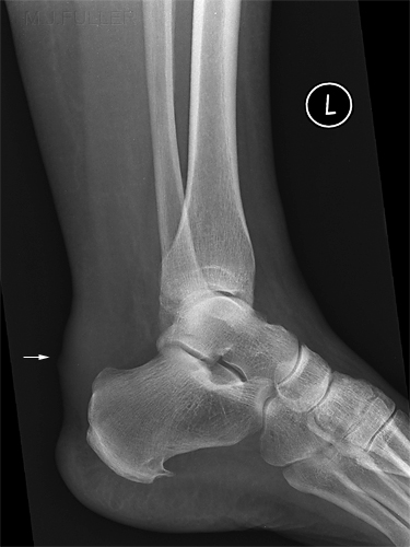
Achille's Tendon Rupture

Soft Tissue Signs in Orthopaedics - wikiRadiography

normal lateral ankle
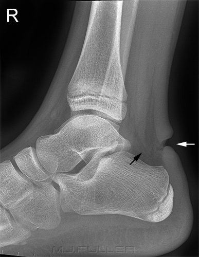
This patient has a ruptured Achilles tendon (white arrow). Note the changes in Kager's Fat Pad (black arrow)
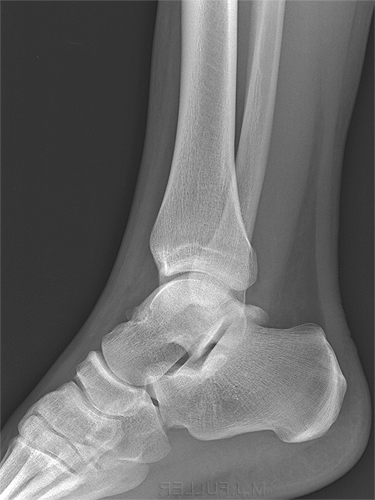
Normal Kager's fat pad with clearly delineated normal Achilles tendon

Complete disruption of the Achilles tendon is most commonly seen in athletes and in men around the age of forty. Achilles tendon rupture can be treated surgically, or by placing the patient in a cast with equinus (marked plantar flexion) for several months. (1)

References
(1) Helms, Clyde A. Fundamentals of Skeletal Radiology.3rd ed. Elsevier Saunders 2005


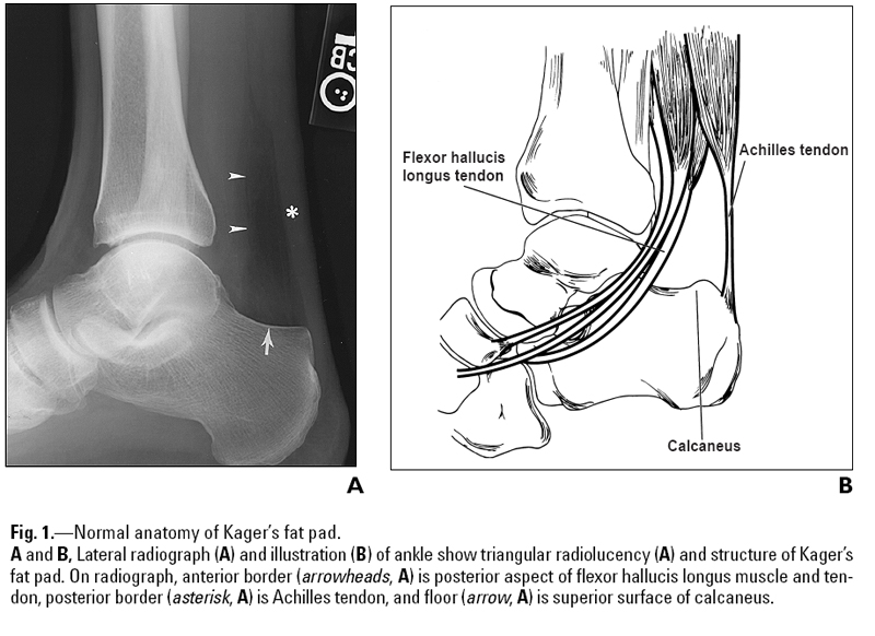
Kager's Fat Pad

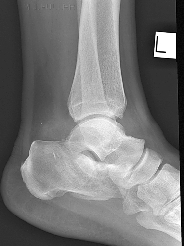
It is not uncommon for ankle injuries to involve Kager's fat pad. A careful examination of the density, shape and borders of Kager's fat pad can provide indicators of bony injury to the ankle. An abnormal Kager's fat pad does not indicate definite bony injury to the ankle.

Soft Tissue Signs in Orthopaedics - wikiRadiography
<a class="external" href="http://www.ajronline.org/cgi/content/full/182/1/147" rel="nofollow" target="_blank">Justin Q. Ly and Liem T. Bui-Mansfield Anatomy of and Abnormalities Associated with
Kager’s Fat Pad AJR:182, January 2004
</a>

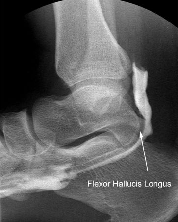
Flexor Hallucis Longus
<a class="external" href="http://radiology.rsna.org/content/236/3/974/F11.expansion.html" rel="nofollow" target="_blank">adapted from
The Flexor Hallucis Longus: Tenographic Technique and Correlation of Imaging Findings with Surgery in 39 Ankles
http://radiology.rsna.org/content/236/3/974/F11.expansion.html
</a>

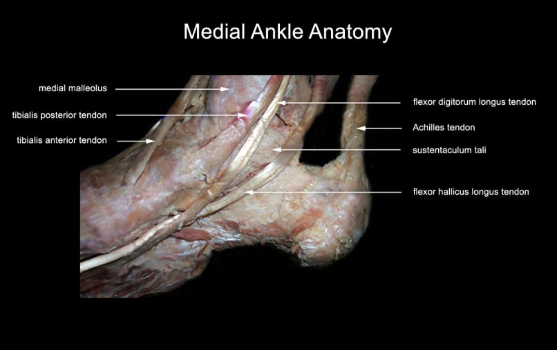
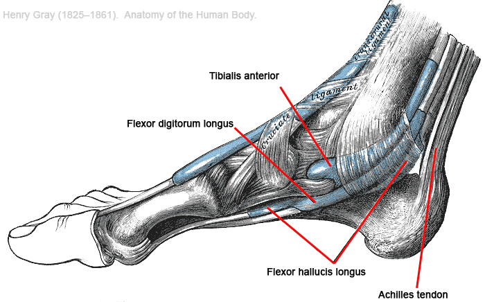
medial ankle anatomy
adapted from
Ithaca CollegeDepartment of Physical TherapyHuman Anatomy Review Sitehttp://www.ithaca.edu/faculty/lahr/LE2000/LE_ankle.html


Further detail of ankle anatomy shown below
lateral ankle anatomy


Abnormal Kagers Fatpadnormal lateral ankle
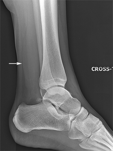
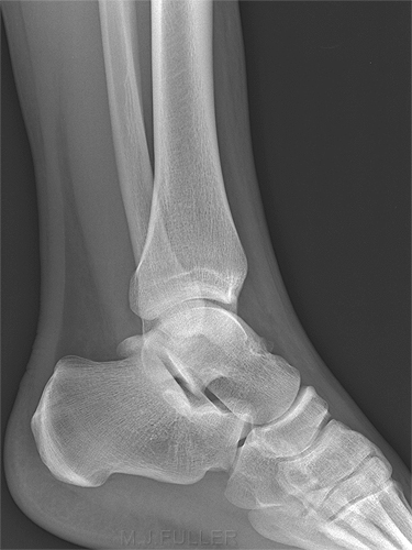
This is a cross-table lateral ankle projection. Note the abnormal fascial plane contour (anterior border of Kager's fatpad). The significance of this finding is that it suggests a significant soft tissue injury and possibly a heightened suspicion of an underlying fracture (although none was demonstrated).Normal Kager's fat pad for comparison


FRACTURED CALCANEUMnormal lateral ankle
This patient presented to the Emergency Department following a fall from a ladder. Note that Kager's fat pad is abnormal showing increased density and indistinct margins. There also appears to be a large ankle effusion. These soft tissue signs should lead you to undertake a careful examination of the bony anatomy. This should be a fruitful exercise- the patient has a fractured calcaneum. Having identified the calcaneal fracture, performing an axial view would be appropriate.Normal Kager's fat pad for comparison


ACHILLES INFECTIONnormal lateral ankle
This 55 year old lady presented to the Emergency Department with a boggy infected area of skin over her Achilles tendon. The patient was referred for ankle radiography with a view to establishing whether there was an underlying osteomyelitis. The infection clearly involves the deeper soft tissues with almost complete obscuration of Kagers fatpad. There is no evidence of osteomyelitis.Normal Kager's fat pad for comparison


Case Study 1

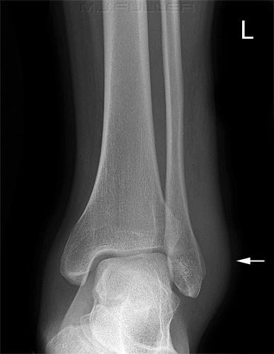
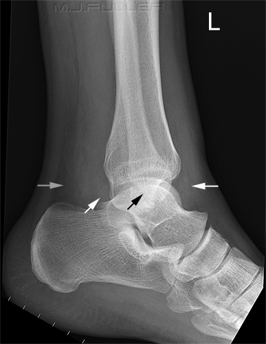
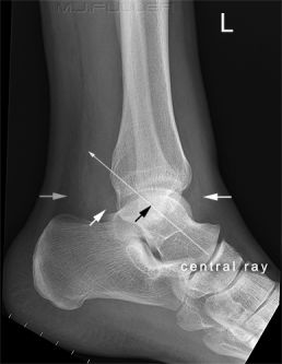
Soft Tissue Signs in Orthopaedics - wikiRadiographyThis patient presented to the ED following a sports injury to the left ankle. On examination, the patient was unable to weightbear (UTWB). Swelling over the lateral malleolus of the ankle was visible clinically and radiographically.
Soft Tissue Signs in Orthopaedics - wikiRadiographyThe lateral view demonstrates

* an ankle effusion- (white arrows)
* abnormal Kager's fat pad (grey arrow)
* suggestion of fracture or accessory ossification centre (os subfibulare)- black arrow


The combination of patient history, clinical signs, soft tissue signs and equivocal evidence of a fracture was sufficient for the radiographer to perform additional views
Soft Tissue Signs in Orthopaedics - wikiRadiographyThe orientation of the possible fibula fracture demonstrated on the lateral projection image suggested that an AP ankle postion with cephalic tube angulation might align the X-ray beam with the plane of the fracture. The strategy was successful demonstrating the disal fibular fracture successfully (left).
lateral ankle

Discussion

This is a good example of the advantages of using a clinical approach to radiography. The radiographer demonstrated a high level of skill in identifying the indicators of an acute bony injury (radiographic and clinical) and performing an appropriate supplementary projection to confirm the presence of a fibular fracture.


Case Study 2

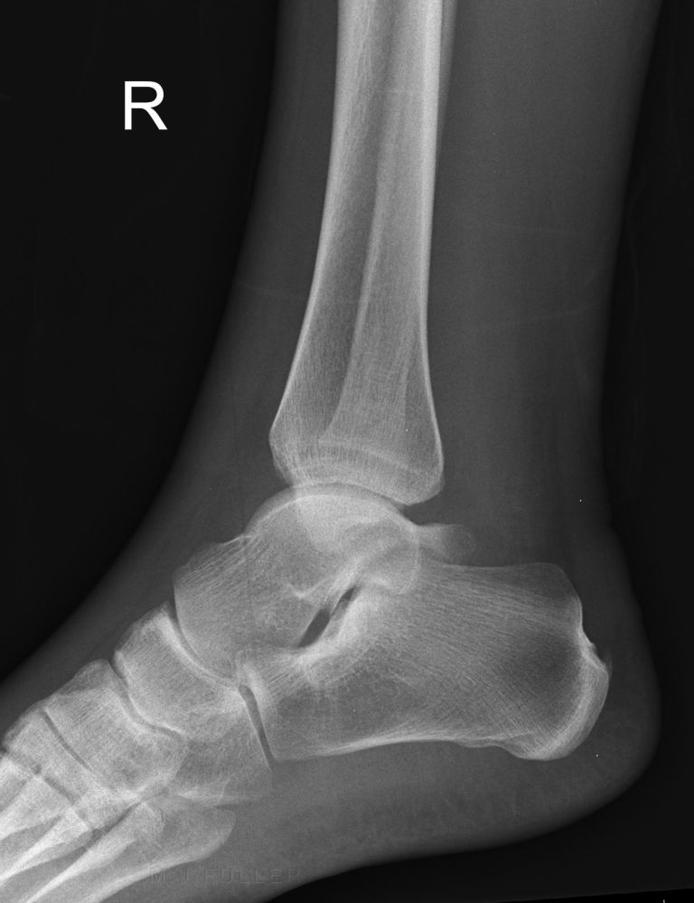
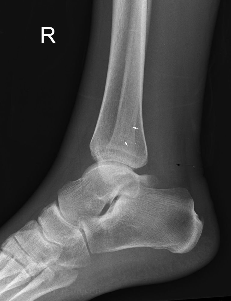
LATERAL ANKLEThis 29 year old male presented to the Emergency Department after rolling his right ankle. The patient was referred for ankle radiography.

The lateral ankle image identifies no convincing bony injury. Kagers fatpad is abnormal. On close examination, there is a suggestion of a fracture of the distal fibula (see below)
lateral ankleThe distal fibular fracture (white arrows) is unconvincing. The Kagers fatpad sign (black arrow) does suggest a significant ankle injury.
Ankle Mortise
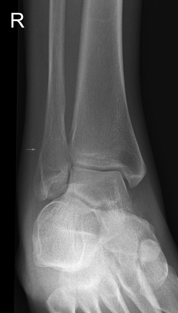
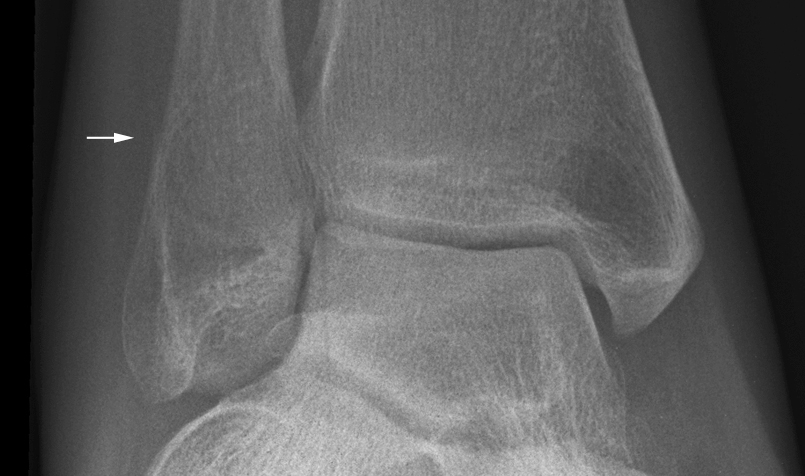
ankle mortiseThe magnified image demonstrates the step in the fibular cortex (white arrow) supporting the diagnosis of distal fibular fracture. There is a poorly demonstrated lucent fracture line (not marked). The diagnosis is also supported by soft tissue swelling over the lateral malleolus.

Discussion

This fibular fracture was identified and red dotted by the radiographer. The abnormal Kager's fat pad drew attention to the possibility of a subtle fracture and the soft tissue swelling over the lateral malleolus supported the diagnosis.


... back to the Applied Radiography home page