Soft Tissue Signs- Skull and Facial Bones

From wikiRadiography
Jump to navigation Jump to search
Introduction

Soft tissue signs of bony injury are commonly demonstrated on skull and facial bone images. This page identifies common soft tissue signs and their significance.


Recent History
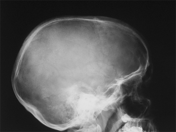
lateral skull radiograph


This is a lateral skull radiograph circa 1990. With film/screen radiography, separate exposures were required to image the skull, facial bones and soft tissues.

This patient was referred to investigate a soft tissue lump on her head. There is no evidence of a soft tissue lump visualised on this image.

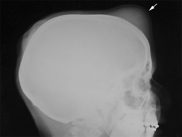
soft tissue lateral skull


The radiographer repeated the view with a soft tissue exposure setting. The soft tissue lump in now clearly visualised (white arrow)


...it wasn't that long ago.... and film/screen radiography is still in use in many parts of the world


Algorithms and Post-processing

If you are using a digital imaging system (DR or CR) correct algorithms and appropriate post-processing are your best friends.

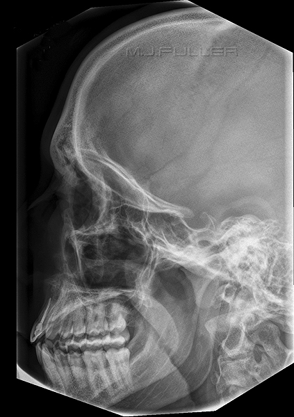
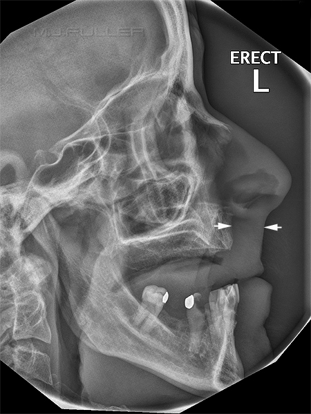
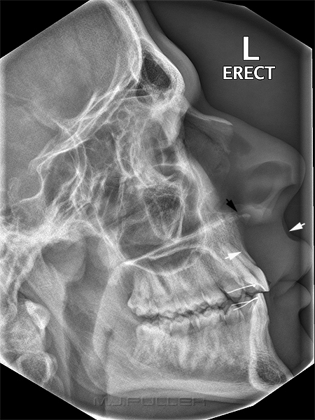
Soft tissue signs are of no utility if you can't see them. Consider the two lateral facial bones images shown below. Both images are taken using the same DR machine.

lateral facial bones 2 lateral facial bones 1
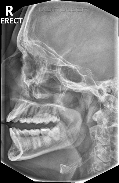
This image looks like a radiograph (slight exaggeration) despite that fact that it was taken using a DR machine. This is the default algorithm/windowing for lateral facial bones for a particular DR machine. The algorithm/windowing does not take advantage of the dynamic range of DR imaging.There is limited soft tissue information and the nasal bones are difficult to see. This image was taken on the same DR machine and using the same defult algorithm/windowing. The radiographer has post-processed the image using the DR software to display soft tissue and bone in the one image. Note that the nasal bones and anterior nasal spine are also clearly demonstrated.


lateral facial bones DR1 lateral facial bones 2
The radiographer has post-processed this DR image to achieve visualisation of the bony and soft tissue anatomy. The soft tissues anterior to the anterior nasal spine can give an indication of underling fracture of the anterior nasal spine. These soft tissues appear normal (arrowed). The radiographer has post-processed the DR image to demonstrate the soft tissues of the face. There is evidence of soft tissue swelling of the upper lip and overlying the anterior nasal spine. The anterior nasal spine appears to be fractured (black arrow). This was confirmed on CT scanning of the skull and face


Case 1
facial bones pa 20 1b

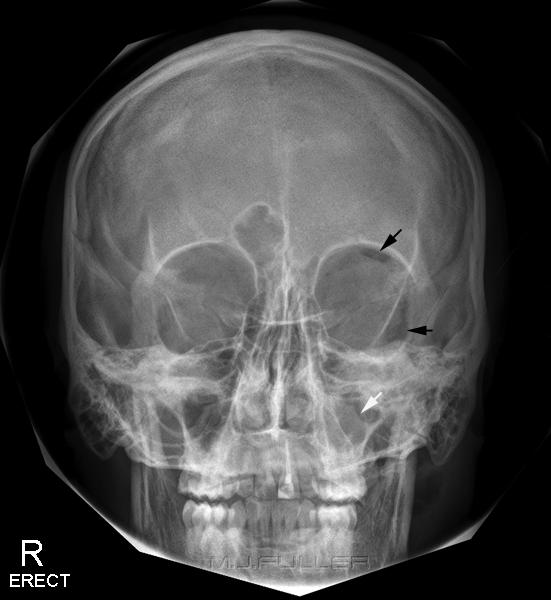
This image was taken using a DR system

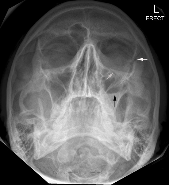
This is a PA 20/25 projection of the facial bones. There is evidence of orbital emphysema (black arrows).

There is opacification of the left maxillary sinus (white arrow)

I don't favour this windowing for facial bones. A facial bone image taken on a DR system should demonstrate all bony structures as well as all soft tissues. This looks like a radiograph rather than a DR image!

CT scanning will often reveal where the subcutaneous air has tracked from. The maxillary sinus or the lateral wall of the ethmoid sinus are commonly involved.

It is not uncommon for patients to be unaware of facial bone fractures until they develop orbital emphysema. This can become apparent to the patient when they sneeze or strain on the toilet.


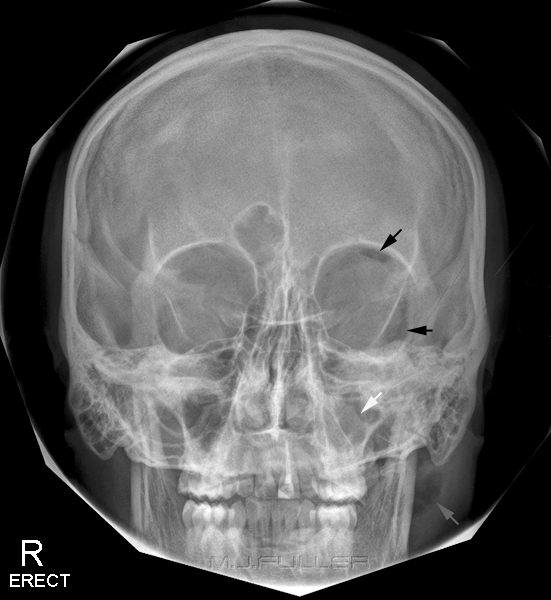
pa 20 facial bones 2


This is the same image which has been post-processed in Photoshop rather than using the DR software- the result is not as good as would have been achieved with the DR software. Despite this limitation, there is now evidence of further subcutaneous emphysema (grey arrow at bottom of image).

There is no justification for creating an image that is high contrast at the expense of its diagnostic value. This may appear self-evident, but I suspect that this "goal-displacement" is more common than we might be prepared to admit.


facial bones om1


The OM view tends to obscure the orbital emphysema. A fracture of the left lower orbital margin is demonstrated (grey arrow). There is soft tissue opacification of the left maxillary sinus associated with the fracture suggesting propulsion of orbital soft tissues into the maxillary sinus. (not arrowed)

The fluid level (blood) is again demonstrated in the left maxillary sinus (black arrow)

The significance of the C1/C2 asymmetry is unknown


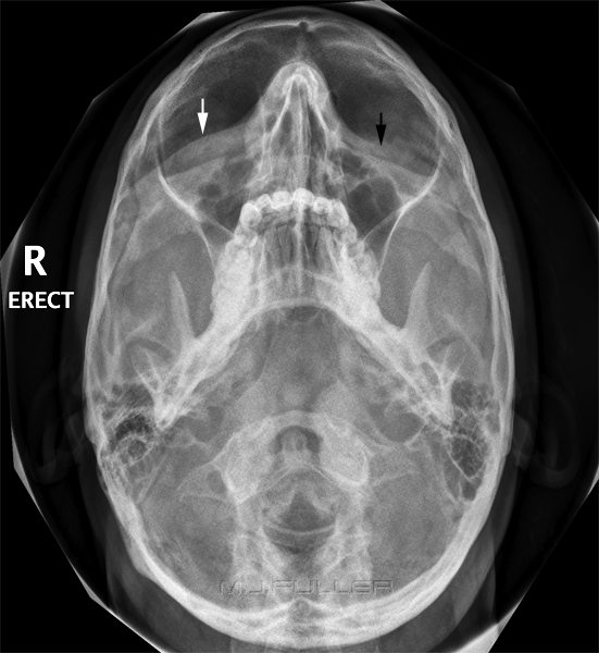
slit basal for zygoma


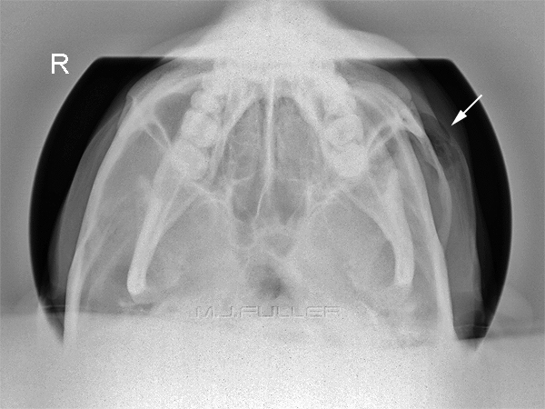
This is a slit basal view of the zygomatic arches. The patient's head has been tilted (laterally flexed) from a true slit basal position to favour demonstration of the affected side (a "teacup view" would arguably have been a better option).

The slit basal image does not demonstrate a fracture of the zygomatic arch.

Subcutaneous emphysema is again demonstrated (arrowed)

Note that the bony anatomy and soft tissues are demonstrated in the same image (once you've used DR, film and CR look like a compromise!)


Case 2

pseudo orbital emphysema


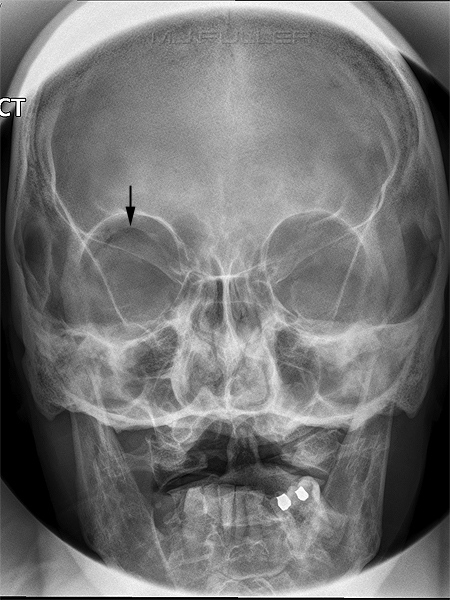
This is a PA 20/25 projection of the facial bones. There appears to be evidence of orbital emphysema (black arrow). This is not however orbital emphysema. This appearance is caused by air trapped beneath the eyelid. This air is typically curved and concave inferiorly. Compare this appearance to the orbital emphysema demonstrated in Case 1 above.


Case 3
facial bones OM 2


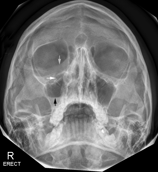
Soft tissue swelling is noted (grey arrow). This soft tissue swelling is projected over the right orbit but is actually overlying the maxillary sinus.

There is evidence of a fracture of the right lower orbital margin (white arrow)

There is a fluid level in the right maxillary sinus (black arrow)


facial bones OM30


The OM30 view similarly demonstrates soft tissue swelling over the right maxillary sinus (white arrow). Compare this soft tissue line with that on the left (black arrow)


Discussion

Facial bone soft tissue signs can indicate underlying bony injury. Good radiographic technique will aid in the visualisation of skull and facial bone soft tissues. The radiographer's goal should be to create an image that displays as much information as possible rather than creating an image that is visually appealing.


... back to the Applied Radiography home page