Soft Tissue Signs- Shoulder

From wikiRadiography
Jump to navigation Jump to search

Introduction

Soft tissue signs can assist in differentiating normal bony anatomy from an acute bony injury. This page considers soft tissue signs associated with the shoulder.


The Normal Humeral Epiphysis

I have seen a normal paediatric proximal humeral epiphysis reported as a fracture on several occasions. In such cases, an examination of the adjacent soft tissues would suggest that no fracture was present.

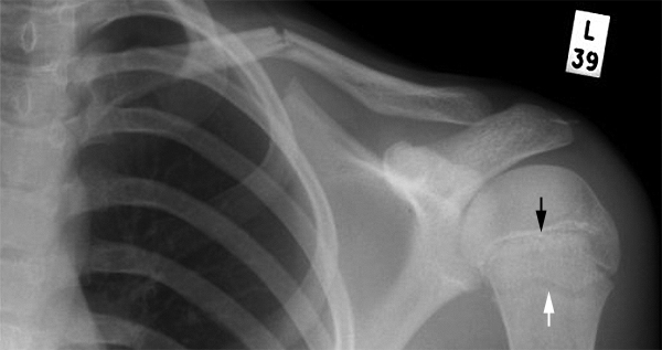
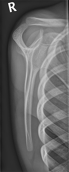
Normal Paediatric Shoulder The arrowed structures are actually a single structure (proximal humeral epiphysis). The proximal humeral epiphysis appears to be two structures for several reasons-

  • The proximal humeral epiphysis is "V" shaped and one side of the "V" is seen en face(ish) and the other side of the "V" is visualised in profile (ish).

  • The other deceptive aspect is that one side of the "V" appears to have a sclerotic margin (black arrow) and the other doesn't(white arrow).
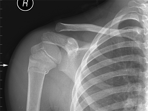
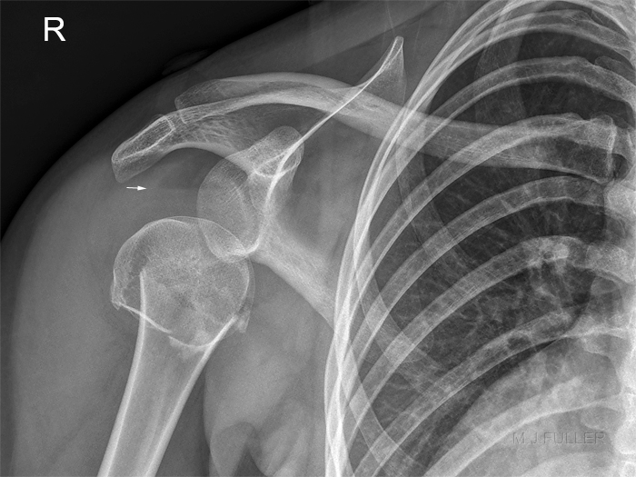
SHI Humerus<a class="external" href="http://www.e-radiography.net/" rel="nofollow" target="_blank">http://www.e-radiography.net/</a> This image can help in understanding the nature and shape of the proximal humeral epiphysis. This patient has a Salter Harris I injury to the proximal humeral epiphysis.


Comment

A normal proximal humeral epiphyses should not be mistaken for a fracture. Examination of the adjacent soft tissues and consideration of the mechanism of injury and clinical signs should further support the diagnosis.


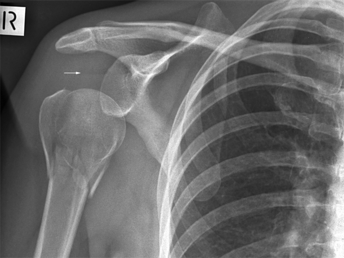
neck of humerus fracture This patient does have a fracture of the proximal humerus. The distinction between the normal proximal humeral epiphyses and the fracture are clearly evident. Note the adjacent soft tissue swelling (white arrow).


Subcutaneous Fat Sign

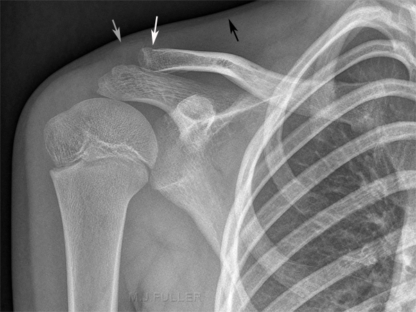
Ap shoulder with Clavicle Fracture There is a suggestion of a step in the cortex of the superior aspect of the distal clavicle (white arrow). There also appears to be an associated lucency in the distal clavicle. There is also a loss of the fat/muscle line adjacent to the cortical irregularity.


Ap Clavicle with fracture The dedicated clavicle image also demonstrates the cortical step and the associated soft tissue sign. Note also the midclavicular canal for the middle supraclavicular nerve

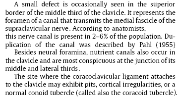
clavicle foramina text
<a class="external" href="http://www.buchhandel.de/WebApi1/GetMmo.asp?MmoId=2998578&mmoType=PDF" rel="nofollow" target="_blank">Source: Koehler/Zimmer's Borderlands of Normal and Early Pathological Findings in Skeletal Radiography, Thieme, 2003, 5th Edition, p307</a>


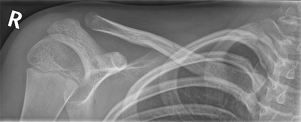
Lateral scapula The lateral scapula image similarly suggests a bony defect in the distal clavicle.


Discussion

The takehome point is that the distal clavicle fracture was subtleish. The associated soft tissue sign provides additional confidence that the defect in the distal clavicle is an acute fracture.


AC joint soft tissue signs
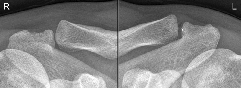
This patient was involved in a highspeed MVA resulting in trauma to his right shoulder. The soft tissues superior to the right AC joint demonstrates swelling and some obliteration of the normal soft tissue planes (compare with left). There is what appears to be a vacuum phenomenon of the left AC joint- this is an interesting observation but is unlikely to be vacuum phenomenon for a variety of reasons, not least of which is that this is the normal shoulder. Note that this is a DR image- the 'vacuum phenomenon' is not a artifact from a fold in the film.


Vacuum Phenomenon
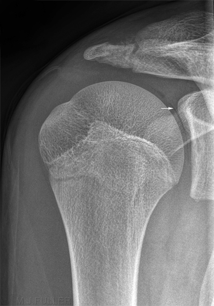
vacuum phenomenonThis is a gleno-humeral joint shoulder image on a 16 year old boy who was squashed between two other players on the sports field. This is his less symptomatic shoulder that was imaged for comparison. The narrow crescent-shaped lucency is gas within the shoulder joint that is associated with a decrease in joint pressure. The appearance is thought to be a possible sequelae of a traction force to the humerus. The traction force results in a pressure reduction in the gleno-humeral joint. The reduction in joint pressure causes nitrogen to be released from solution. What you are seeing is a part of the articular cartilage of the gleno-humeral joint contrasted by gas.

Negative Pressure in Joint

The glenohumeral joint is normally bathed in a small amount (less than 1 mL) of free synovial fluid. Under normal conditions, the intra-articular pressure is negative, which creates a relative vacuum that resists bony translation

Caused by Arm Pulling

Caused by pulling on the arm and thereby decreasing the pressure of the synovial fluid

Nitrogen Release

The decrease in pressure causes nitrogen to be released from solution

Soft Drink Analogy

This is akin to releasing the pressure in a soft drink by releasing the cap


Lipohaemarthrosis
NOH fractureLipohaemarthrosis refers to a condition in which an intra-articular fracture has allowed fat to be released from bone marrow into the joint. As fat is less dense than blood, the fat 'floats' on the blood in the joint and results in an fat/blood "fluid level". This is akin to the air/fluid level seen on erect abdominal plain film images- the difference being that there are two immiscible fluids (fat and blood) rather than a gas and a fluid (air and water). With horizontal beam radiography, a fat-fluid level is detected due to differences in attenuation of these two substances.

Lipohaemarthrosis is one of the few reasonably reliable soft tissue signs of fracture. Lipohaemarthrosis is usually associated with intrarticular knee fractures but can occur (in theory) in just about any synovial joint and is occasionally seen in the shoulder joint.

Where a shoulder fracture is subtle, a lipohaemarthrosis is a useful soft tissue indicator of intra-articular fracture.

A lipohaemarthrosis will only be seen when patients are imaged with a horizontal beam (i.e. erect).

There is inferior displacement of the humeral head on the glenoid. This appearance (pseudosubluxation) is commonly seen in association with fracture and suggests joint effusion.
SHOULDER LIPOHAEMARTHROSISThis patient has a neck of humerus fracture, pseudosubluxation of the GH joint, and lipohaemarthosis.


... back to the Applied Radiography home page