Small Bowel Obstruction
Small bowel obstruction (SBO) is a common abdominal pathology. This page examines radiographic techniques and pathological appearances of plain film small bowel obstruction. Before reading this page, it would be advisable to read the page on terminology and the page on differentiating small and large bowel appearances on plain film.
Definition of Small Bowel Obstruction (SBO)
Radiographic TechniqueSmall bowel obstruction refers to any condition where there lumen of the small bowel is obstructed. The obstruction may be intrinsic (as with intussusception) or extrinsic (as with abdominal adhesions). A small bowel diameter on plain film greater than 30mm is considered dilated.
Appropriate views for an acute small bowel obstruction should start with a supine abdominal plain film X-ray. It could be argued that all acute abdominal imaging should include an erect AP/PA chest view to demonstrate pneumoperitoneum. There is also the possibility that the abdominal symptoms are misleading.
The erect abdominal plain film should be undertaken to clarify the appearances on the supine image, or where there is a reasonable expectation of additional relevant findings (see discussion below and here). The erect abdominal plain film should not be performed in isolation under normal circumstances.
Where there are equivocal findings on the supine image, a repeat supine abdominal plain film image can be very useful. It should be remembered that an abdominal image provides a snapshot of the bowel at a particular point in time. If there is an equivocal or suspect appearance, a repeat view taken as little as 10 minutes later will often confirm an appearance as being pathological rather than a normal transient chance appearance.
A rarely utilised projection in the acute setting is the prone abdominal view. The advantage of the abdominal plain film with the patient in the prone position is that the dependent and non-dependent parts of the bowel are reversed. The disadvantage is that the patient is unlikely to find the position comfortable.
The left lateral decubitus view is a suitable alternative to the erect abdominal view. A right lateral decubitus or supine cross table lateral decubitus should also be considered in appropriate circumstances.
A tangential view of external hernias can reveal incarcerated small bowel (see examples on this page).
The Erect Abdominal Film in Small Bowel Obstruction
The difference between the supine abdominal plain film and the erect abdominal plain film has plenty to do with gravity and not much else. Not to put too fine a point on it, the AP supine abdominal plain film and the erect AP abdominal plain film are projections of the same anatomy and are even under the same gravitational forces. The difference lies in the fact that a contained air/fluid filled structure will have an interface that is imaged en face in the supine projection and in profile in the erect projection.
There are, of course, other more subtle benefits of the erect plain film as follows
- potential to include all of the upper abdomen (centre slightly more superiorly)
- in a gasless obstruction, loops of fluid filled small bowel can be seen to drop under the effect of gravity
- possibility of demonstrating a string-of-pearls sign
- others I'm sure
The PosterI have placed a poster on the wall of the X-ray viewing area in the Emergency Department as shown below
This is a quotation from the abdominal plain film bible by Stephen R. Baker. His book titled The Abdominal Plain Film is out of print but second hand copies are available from Amazon. In my opinion it's well worth the money. We have a copy in our Radiology library but it is continuously out on loan.
Abdominal plain films are not necessarily either normal or abnormal; this is not a dichotomy- it's a continuum. If you are one of those people that likes to categorise things, you could think of them as being;
normal -------------> probably normal ----------> suspect ------->probably abnormal-----> definitely abnormal


The Role of CT
CT is the gold standard of diagnostic imaging in patients with acute abdominal symptoms. Whilst a plain abdominal film can suggest a diagnosis of small bowel obstruction, CT is more likely to reveal the cause and site of obstruction.
Clinical Presentation of SBO
abdominal pain
squealing bowel sounds (early obstruction)
no bowel sounds (bowel wall muscular exhaustion)
rapid onset of nausea and vomiting
belching
abdominal rigidity
abdominal swelling
Causes of Small Bowel Obstruction
Treatment of SBO
- Adhesions
- Neoplasms
- Hernias-external, internal
- Crohns
- Other
The most common cause of small bowel obstruction in the western world is from adhesions secondary to abdominal surgery. An adhesion is an abnormal band of tissue which can indent/deform the small bowel causing obstruction.
It is useful for the radiographer to establish with the patient whether they have had abdominal surgery in the past - this may assist in interpretation of the plain film abdominal image. Furthermore, it is helpful to ask the patient if they have had multiple episodes of SBO from adhesions in the past.
Consider any abdominal wall asymmetry as a potential hernia or other pathology. Radiographers have demonstrated umbilical and incisional hernias only because they have noticed the patient had an unusual abdominal wall asymmetry.
(Some texts will show hernias as the second most common cause of SBO)
The treatment of SBO will vary with the circumstances of individual cases. Insertion of a naso-gastric tube into the patient's stomach is a common treatment. Some patients are treated conservatively to see if the SBO will resolve spontaneously. Gastrografin has also been used as a therapeutic agent in SBO. Surgical intervention is sometimes required.
Diagnostic Accuracy
The abdominal plain film is a blunt diagnostic tool. It has been likened to taking a patient's temperature- an abnormal plain film appearance suggests that there is abdominal pathology present much the same as a high temperature suggests that a patient has an infection.
The 3,6,9 Rule
There is a good report on a study investigating the accuracy of the abdominal film in the diagnosis of SBO here <a class="external" href="http://www.ajronline.org/cgi/reprint/188/3/W233" rel="nofollow" target="_blank">http://www.ajronline.org/cgi/reprint/188/3/W233</a>
The maximum diameter of the bowel is shown below
Maximum Normal Diameter small bowel 30mm large bowel 50-60mm caecum 90mm
The 3,6,9 rule is a very useful guide to determining when the bowel is dilated. It can also be useful in distinguishing between small and large bowel. For example, if the small bowel measures 90mm in diameter it is probably not small bowel.
Geometric Magnification Issues
The 3,6,9 rule is for uncorrected measurements. The error associated with an uncorrected measurement is usually not a problem. Where it can be a problem is in morbidly obese patients where the small bowel is situated close to the LBD/focal spot.
If you perform erect abdominal images PA rather than AP you may identifying small bowel affected by geometric enlargement demonstrated on the supine image

Small Bowel Appearances Indicating SBO
- small bowel dilated over 30mm
- multiple air/fluid levels in small bowel
- stretch/slit sign
- string of pearls sign
- coiled spring sign
The important finding in SBO is a change in calibre of the small bowel. If the SBO is sufficiently obstructed, the small bowel proximal to the obstruction will dilate. Small bowel with a diameter greater than 30mm is considered to be dilated. Small bowel can dilate up to around 50mm. If the small bowel has a diameter of 70mm or greater it probably isn't small bowel.
The stretch sign or slit sign in which a slit of air caught in a valvulae is characteristic of SBO.
The erect abdominal image will show multiple air/fluid levels in dilated small bowel in patients with SBO.
<a class="external" href="http://www.dhmc.org/dhmc-internet-upload/file_collection/10.20.04+-+Bowel+Obstruction.pdf" rel="nofollow" target="_blank">http://www.dhmc.org/dhmc-internet-upload/file_collection/10.20.04%20-%20Bowel%20Obstruction.pdf</a>This is an example of SBO at surgery. Note that the bowel is dilated proximal to the point of obstruction and collapsed distal to the point of obstruction.

Can a SBO be Differentiated from an Ileus on Plain Film?
The hallmark of small bowel obstruction is the presence of gaseous loops of small bowel which are distended over 30mm. The absorptive capacity of the small bowel is so great that even extreme amounts of air swallowing will not distend normal small bowel.
The presence of dilated loops of small bowel is not a guarantee that the patient is obstructed. Correlation with patient history and clinical signs can assist in arriving at a more specific diagnosis. The difficulty in differentiating obstruction from ileus has led some radiologists to use the blanket term "motility disorder" when describing dilated loops of bowel.
There is a very good discussion on obstruction vs ileus in paediatrics here <a class="external" href="http://www.hawaii.edu/medicine/pediatrics/pemxray/v3c18.html" rel="nofollow" target="_blank">http://www.hawaii.edu/medicine/pediatrics/pemxray/v3c18.html</a>
Note the logical process of arriving at a probable diagnosis.
Small Bowel Obstruction- Establishing a level of Obstruction
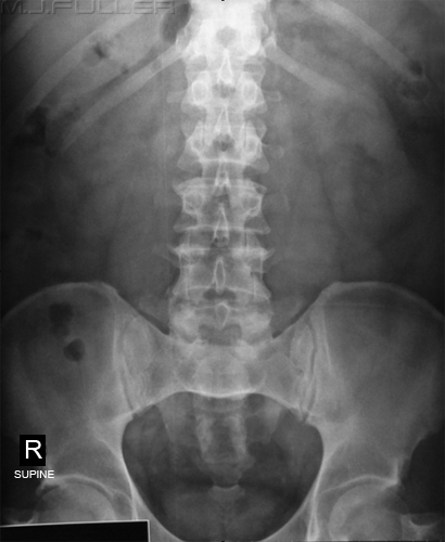
This patient has a solitary loop of air-filled dilated small bowel in the left upper quadrant. This loop of jejunum has a coiled spring appearance that is associated with SBO.
Given that there is only one proximal loop of dilated small bowel it would appear reasonable to assume that the obstruction is very proximal. It is possible that the obstruction is more distal than is suggested by this solitary loop of dilated small bowel. What is difficult to appreciate is that there may be multiple fluid-filled dilated loops of small bowel which are not clearly visualised.
There is evidence of a collapsed large bowel suggestive of SBO

LBO Posing as an SBO
At a cursory glance this patient appears to have a SBO. On closer examination, the prominent air-filled loops of small bowel in the LUQ (white arrow) have the features of ileum rather than jejunum. Also, the caecum appears unusually large and there appears to be a sudden change in calibre in the large bowel at the level of the hepatic flexure(black arrow). This was reported as a SBO. An alternative explanation is that the large bowel is obstructed at the level of the black arrow. This would account for the dilation of the caecum. A LBO in patients with incompetent ileocaecal valves can mimic a SBO. The ileal loops may have been displaced by the enlarged caecum or they may be effaced jejunal loops.
Note that the patient has the reliably unreliable sign of gas in the rectum!
This is a barium enema on the same patient. Note the tight apple-core lesion at the level of the hepatic flexure (white arrow)
Approximately a quarter of patients have an incompetent ileocaecal valve.


The Normal Small Bowel
One of the mantras of image interpretation is that you will not be able to recognise an abnormal appearance if you are not able to identify a normal appearance. This is highly relevant to the small bowel which demonstrates a highly variable normal range of appearances.
Normal Gasless Small Bowel
Normal Air-filled Small Bowel
This patient appears to have a gasless small bowel. Fluid-filled loops of small bowel are present but are never as well visualised as air-filled loops.
There is debate regarding what is a normal amount of small bowel gas. Gas enters the small bowel during eating and drinking. Some people habitually air-swallow while others air-swallow at times of stress or when they are in pain. If the patient is air-swallowing at a higher rate than the small bowel can absorb the air, it will be visualised in the bowel. Some texts suggest that up to seven fluid levels in the small bowel on an erect abdominal film can be normal. Most texts quote a more conservative figure.
I have heard it said that any air visualised in the small bowel is abnormal. I find this assertion hard to support.
Don't confuse gasless with featureless. A featureless abdomen can be a result of tumour or ascites. This patient has a gasless rather than featureless abdomen. Note that the renal, liver, psoas muscles and urinary bladder outlines are visualised
Note that this abdominal film is not guaranteed to be normal. It could represent an early gasless small bowel obstruction. Clinical correlation is required.

This is an AP lumbar spine image on a patient who presented with back pain. It would be reasonable to assume that this patient is unlikely to have acute abdominal pathology (although not completely excluded). The appearance of the small bowel visualised in the left iliac fossa is a result of normal air swallowing (white arrow). The bowel diameter has been measured at 30mm which is the upper limit of normal. This patient is likely to be in pain and is therefore more likely to air-swallow resulting in this appearance.
The appearance has been likened to crazy paving or the pattern on a giraffe. It appears as an interlocking, random, tessellated pattern.
Crazy Paving Giraffe!


Minimally Dilated Small Bowel
The small bowel demonstrated in this image is minimally dilated (36mm). There is evidence of loss of the normal random tessellated pattern associated with undilated small bowel. Instead, the bowel is showing signs of a pattern which is more organised rather than random. There are, for example, multiple loops of small bowel which have become aligned/parallel.
This appearance may represent an early small bowel obstruction or a partial small bowel obstruction. Clinical correlation and an erect film may be very helpful in determining whether the appearance is pathological.
This appearance is not typical of generalised adynamic ileus although this cannot be excluded.

Gasless Small Bowel Obstruction
The coiled spring appearance only occurs in the dilated air-filled small bowel. It also is most noticeable in the jejunum where the valvulae conniventes are closely spaced.


You could be forgiven for thinking that this patient has been drinking dilute gastrografin. This appearance is a gasless small bowel obstruction and the opaque looking small bowel loops (white arrow) are filled with normal small bowel content, rather than gastrografin. If you compare this image with the gasless small bowel image above you can see that this small bowel is significantly more prominent.
There can be difficulty in distinguishing an early gasless small bowel obstruction from a normal appearance of the small bowel in someone who has just eaten a large meal.
Clinical correlation and follow-up imaging will usually provide confidence in the diagnosis.
The large bowel is not clearly visualised suggesting that it may be collapsed.

String-of-Pearls Sign
Slit/Stretch Sign
The curvi-linear arrangement of air bubbles visualised on this image is known as the string of pearls sign. The appearance is considered to be diagnostic of obstruction (as opposed to ileus) and is caused by small bubbles of air trapped in the valvulae of the small bowel. Source:Abdominal radiology [Hardcover]James J. M.D. McCort , Robert E. M.D. Mindelzun, Robert G. M.D. Filpi , Charles M.D. RennellWilliams and Wilkens 1981p 117,148,151.
“The “String-of-pearls’ sign [is]… almost always indicative of intestinal obstruction and is one of the few situations in which an upright or decubitus film of the abdomen contributes crucial information about small bowel obstruction.”
Appleton & Lange, 1990, p156
A similar appearance is sometimes seen in the large bowel but can usually be differentiated by the fact that the gas bubbles are larger and have flat under-surfaces
There is an excellent article on string of pearls sign <a class="external" href="http://radiology.rsnajnls.org/cgi/reprint/214/1/157.pdf" rel="nofollow" target="_blank">here</a>


The Large Bowel String of Pearls Sign
This patient has a small bowel obstruction. Apart from the solitary air-filled dilated central loop of small bowel, there is also evidence of slit sign or stretch sign (white arrows).
Slit sign is a result of small amounts of air caught in the valvulae of fluid-filled bowel. The subtle fluid filled loops of small bowel and the slit sign are highly suggestive of small bowel obstruction. This appearance is deserving of an erect abdominal projection. This patient had one of the best string of pearl signs you will ever see!Abdominal radiology [Hardcover]James J. M.D. McCort , Robert E. M.D. Mindelzun, Robert G. M.D. Filpi , Charles M.D. RennellWilliams and Wilkens 1981p 117,148,151.


The large bowel has its own version of the small bowel string of pearls sign. Because the plicae semilunaris of the large bowel are larger than the valvulae of the small bowel the pockets of air tend to be larger. Also, because they are larger in the large bowel, surface tension is unable to make them round- instead they tend to have a flat underside. They look more like a string of air-fluid levels.
One of the functions of the large bowel is to absorb water from the faecal content. The faeces should not be able to form an air/fluid level by the time it gets to the splenic flexure. An extensive arrangement of these small air/fluid levels in the large bowel may indicate that the patient has diarrhoea.

Multiple Air-fluid Levels
Tangential Views of External Hernias
This is an erect PA abdominal image. The PA erect abdominal projection has several advantages:
- There is potential for the patient to hold onto the erect bucky making them feel safer and more secure
- It is easy to adjust the tube and bucky position to a PA chest position
- The anteriorly sited small bowel and transverse colon will be close the bucky/IR reducing the potential for geometric magnification
There are dilated loops of small bowel and multiple air-fluid levels. This appearance is characteristic of small bowel obstruction. Additional confidence that the appearance is caused by small bowel obstruction is afforded by the string of pearls sign (white arrow).
The dilation of the small bowel stimulates the mucosa to secret fluid. A quantity of fluid and ingested gas in a contained structure and a horizontal beam are all the necessary requirements for an air-fluid level on plain film
The appearance of multiple air-fluid levels on erect abdominal film is sometimes referred to as a step ladder sign. It has also been suggested that uneven levels in a bowel loop (i.e. more fluid on one side of the loop than the other) is diagnostic of SBO rather than ileus. This is refuted by some authors.

The Gas in the Rectum "Conspiracy"
This patient presented with a history consistent with small bowel obstruction. The patient also reported that a lump had appeared on her anterior abdominal wall.
The radiographer has undertaken a lateral abdominal plain film in the supine decubitus position. This position may be preferable to the erect lateral position shown below. The advantage of the supine decubitus position is that there may be a greater chance of air entering the herniated bowel because it is the least dependent part of the bowel in the supine position. Gasless incarcerated small bowel in an external hernia may be difficult to visualise.
An aluminium filter covering the herniated bowel is very useful when imaging abdominal hernias in a tangential projection. Also, DR is better than CR, and CR is better than film/screen.This patient presented to the Emergency Department with a similar history. The radiographer has performed an erect lateral abdomen revealing an umbilical hernia containing small bowel and an air/fluid level.
The herniated abdominal contents may not appear to contain bowel if the bowel is gasless.


The presence of gas in the rectum is widely considered a useful indicator to exclude bowel obstruction. The reasoning is that if the bowel is obstructed, there should be no passage of gas to the rectum. This sign is unreliable. The bowel is often partially obstructed, allowing the passage of bowel contents past the level of the partial obstruction. More importantly, the large bowel produces its own gas through fermentation processes. Even in cases of complete obstruction, gas in the rectum may persist for several days. A lack of gas in the rectum is worthy of consideration, but is not a reliable sign of bowel obstruction. Equally, gas in the rectum does not exclude bowel obstruction. A collapsed large bowel is arguably a more reliable sign of bowel obstruction.
Limited Gastrografin Follow-through
The limited gastrografin follow-through has become a popular examination in recent years. The objective is simply to establish if orally administered gastrografin is propelled throughout the small bowel to the large bowel. An added benefit of gastrografin is that it is a <a class="external" href="http://www.ncbi.nlm.nih.gov/pubmed/12131078" rel="nofollow" target="_blank">hyperosmolar</a> water-soluble contrast medium. Its hyperosmolar properties can have a therapeutic as well as diagnostic benefit.
One of the difficulties with the limited gastrografin follow through examination in patients with suspected SBO is that it can be self-defeating. That is to say, the aim of the study is to examine the gastrografin as it passes through the bowel...and the indication for the examination that there is something causing stasis of bowel contents.
It is not uncommon for the gastrografin to become so dilute that it is barely detectable. It is possible to add a small quantity of barium (say 10 mls) to the gastrografin as a trace contrast medium. The barium tends to leave a trail of where it has been. The argument against this is that the barium will become an irritant if it enters the peritoneal cavity through a bowel perforation. The counter argument is "... not as much as the bowel contents".
The limited gastrografin follow through is a popular study with the surgeons. The expectation is that if the gastrografin makes it to the caecum, surgical treatment of the SBO will probably not be required or not required urgently.

Generalised Adynamic Ileus
The bowel could reasonably be said to be a very sensitive organ. It has a propensity to stop functioning with little provocation. Amongst the possible causes are infection(anywhere), abdominal inflammation, chemical/pharmacological causes and trauma.
Abdominal surgery commonly results in generalised adynamic ileus in which the bowel is temporarily non-functioning. This typically manifests on day 4 post-op. In response, the patients are often referred for abdominal plain film imaging to rule out bowel obstruction.
The appearance of generalised adynamic ileus is quite characteristic. The large and small bowel are extensively airfilled but not dilated. I have heard this described as the large and small bowel "looking the same".

Case 1
This 70 year old lady presented to the Emergency Department with abdominal pain and distension. She reported a history of bowel cancer.
There is stretch sign indicating distended fluid-filled small bowel strongly suggestive of small bowel obstruction.
The large bowel contains very little faeces and is largely gasless.There erect abdominal plain film demonstrates string-of-pearls sign which is considered pathognomonic of small bowel obstruction.


Further Reading
Unless I am mistaken, this is the best textbook ever written on the abdominal plain film. It is well referenced and written in a readable style. It is out of print.
I have an older version of this book which I purchased second hand through Amazon. It was well worth the money!
I have also added the follwoing text onto my wishlist...
Abdominal radiology [Hardcover]
James J. M.D. McCort , Robert E. M.D. Mindelzun, Robert G. M.D. Filpi , Charles M.D. Rennell
Williams and Wilkens 1981

Summary
Small bowel obstruction is a common pathology. A knowledge of the patterns of normal small bowel and small bowel obstruction will assist in the interpretation of plain film findings. Importantly, correlation with clinical findings, patient history, and other test results can give an equivocal appearance additional meaning. Finally, the radiographer's knowledge of normal and pathological appearance will allow him/her to make an informed decision as to when supplementary views are justified and what views would be most suitable.