Introduction
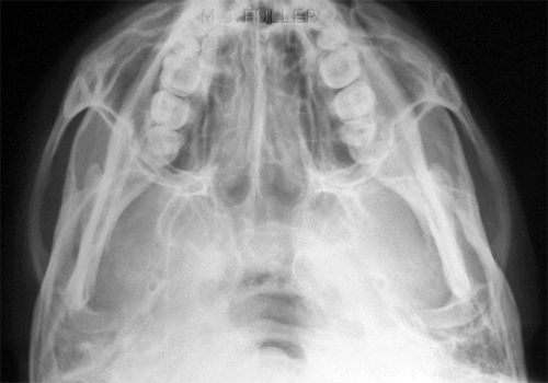
The slit basal view of the zygoma is an effective view for demonstration of fractures of the zygoma. The radiographic technique is not as difficult to master as it might appear. One of the advantages of the slit basal and teacup projections is that they can provide a true representation of the degree of depression of a zygoma fracture- this is likely to be influential in the decision to perform a surgical zygoma elevation operation.
Radiographic PositioningSlit Basal
| 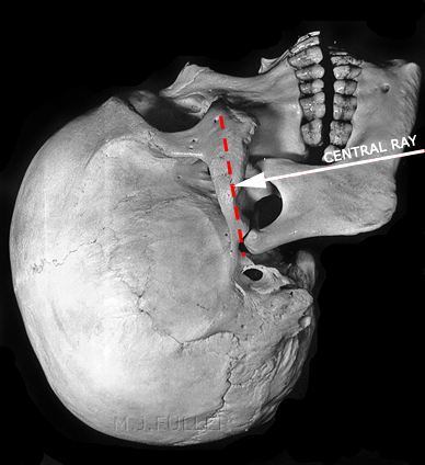 |
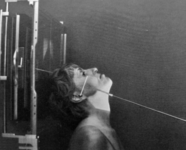
| The patient is seated with their back to the wall bucky/IR. The mid-saggital plane of the skull is at right angles to IR and the beam is angled at 90 degrees to the zygomatic arch. Centre to the level of the middle of the zygomatic arch in the mid-saggital plane. | This view can be achieved with the patient in the erect AP or supine position. A sponge under the patient's shoulders can assist the patient to achieve greater neck extension. |
Teacup View
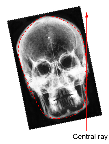 | 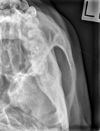 |
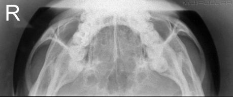
When imaging one side only, the positioning is similar to the slit basal view. The following should be noted
- collimate to include the anatomy of interest
- centre to the zygomatic arch of interest
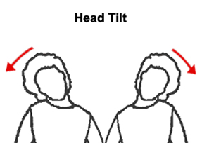
- laterally flex the patient's head away from the side of interest
- radiographers are known to confuse which direction to laterally flex the patient's neck for a teacup view. It may be of assistance to think of the patient as having a face that is slightly wedge-shaped.
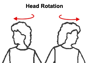
- To achieve a teacup view position, the patient's head is laterally flexed not rotated.
| 
The teacup view derives its name from a likeness to the handle of a teacup. |
To achieve a teacup view position, the patient's head is laterally flexed/tilted not rotated.
Notes
- The slit basal and teacup views are contraindicated in a patient that may have a cervical spine injury.
- There is considerable flexibility in the beam angulation relative to the zygomatic arch. If the beam is not perfectly directed at 90 degrees to the zygoma, the results are likely to still be acceptable.
- You will learn to read a patient's zygomatic arches- patients with chiselled features and "high cheek bones" tend to have zygomatic arches that sit proud of the skull and are therefore easy to image. Some patients have a facial bone structure that does not accommodate successful application of the slit basal technique.
Beam Collimation
A round cone can be employed to good effect when performing slit basal radiography of the zygomatic arches.
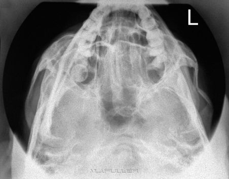
|
Slit basal projection with round cone demonstrating a left comminuted and depressed fracture of the zygoma |
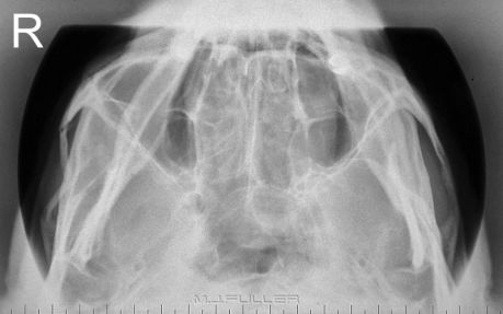
|
same again |
Correcting Positioning ErrorsCase 1
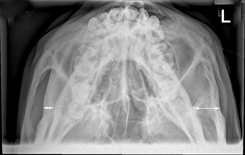 | This patient presented to the Emergency Department following facial bone trauma. The radiographer has performed a slit basal view of the zygomatic arches. The patient's head is laterally flexed to the left. This positioning error tends to favour demonstration of one zygoma (in the case the right zygoma) at the expense of the contra-lateral side.
Rather than repeat the slit basal view, the radiographer has performed a teacup view of the left zygoma. |
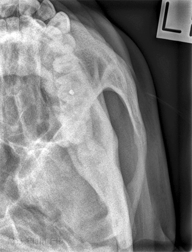 | The teacup view demonstrates the left zygomatic arch successfully. |
Case 1 Discussion
This case raises a few questions. If the patient has sustained a blow to the left zygoma alone, why not perform a teacup view of the affected side only? Alternatively, if your departmental routine is to image both zygomas, why not laterally flex/tilt the patient's head to favour the zygoma of interest? Irrespective of your answers to these questions, I would suggest that radiographers should be mindful of these options, and, if your scope of practice allows you to, modify the series to suit the clinical situation.
Case 2
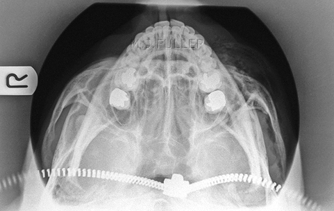 | When performing a slit basal view with the patient in the supine position, it is very easy to overlook potential image artifacts from items on the patient's chest.
This zipper artifact could be avoided by removing the artifact or by extending the patient's head further and angling the X-ray beam more to match. |
Case 3
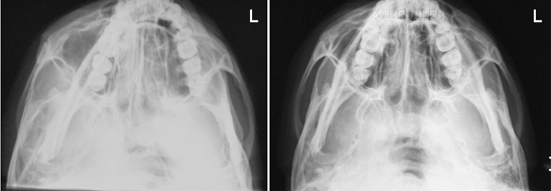 |
| The first attempt favours the left zygoma at the expense of the right. The right image shows correction of the left lateral flexion/head tilt positioning error. |
Case 4
This patient presented to the Emergency Department after a direct blow from a baseball to the right TMJ/zygoma. There was a clear rounded swelling at the site of impact. The patient was in great pain and clearly distressed. He was still able to talk but had limitations of his jaw movement. The patient was referred for TMJ and facial bone radiography. He had received no pain relief at the time of the X-ray examination. Note the sequence of imaging, the use of supplementary views, and the logical path the radiographer took based on the radiographic findings combined with clinical signs and patient observation.
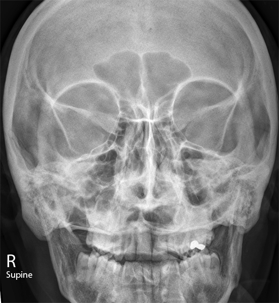 | The patient's degree of distress and lack of pain relief suggested to the radiographer that erect imaging of the facial bones was risky in terms of the patient fainting. Facial bones can be imaged supine successfully.
This is a supine AP 25 facial bones. No significant abnormality was detected on this image |
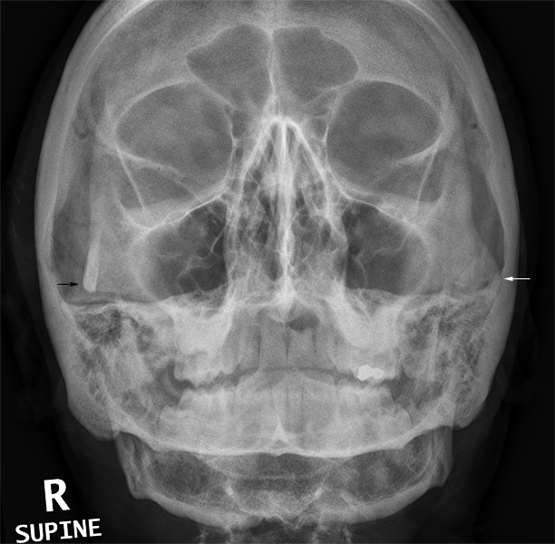 | The supine OM 30 view is undertilted. The projection is actually MO (the beam passes from the mental point of the mandible to the occiput) but common practice is to describe the position as OM. IT is not unusual for the combination of patient neck extension and tube angle to produce an under-tilted OM image. This is a legacy of the additional difficulty of the patient achieving the OM position whilst supine (that's not to say it can't be done).
The radiographer noted the asymmetry of the zygomatic arches (arrows) |
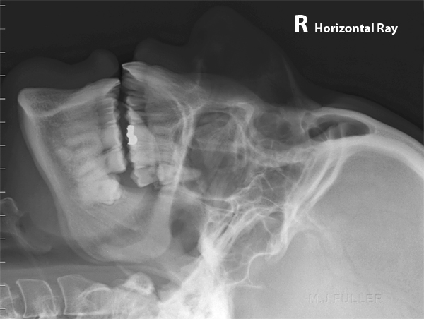 | The lateral facial bones were performed with a horizontal beam. No significant abnormality was detected. |
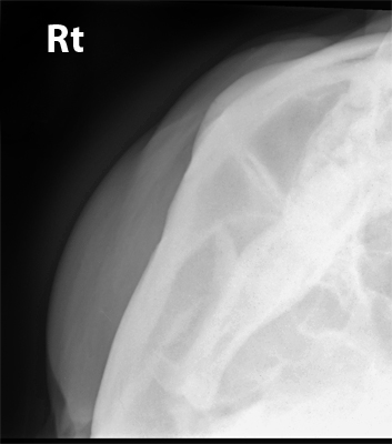 | The routine series for facial bones included a slit Townes or slit Basal for zygomatic arches. The radiographer chose to perform the slit basal in preference over the slit Townes.
The slit basal is not a difficult technique in a supine patient. The patient position is not that dissimilar to that which was employed for the supine OM view. A non-grid cassette was placed above the patient's head in a slightly off-vertical position. The X-ray tube was positioned over the patient and directed with a slight downtilt (10-20 degrees depending on the degree of neck extension) and centred on the zygoma.
This patient had a clearly defined unilateral injury. It is prudent in such cases to perform a teacup view rather than a slit basal for zygomas. The teacup view has the advantage of not irradiating the normal side unnecessarily, and the patient's head can be laterally flexed/tilted to favour demonstration of the affected side by bringing the zygomatic arch further into profile.
The technique failed to provide an image of the affected zygoma. This was surprising given that the patient appeared to have prominent zygomas and the teacup view's advantage over the slit basal is that it can demonstrate a zygomatic arch in a patient that does not have prominent zygomatic arches. Consideration was given to the possibility that the patient had a depressed fracture of the right zygoma. |
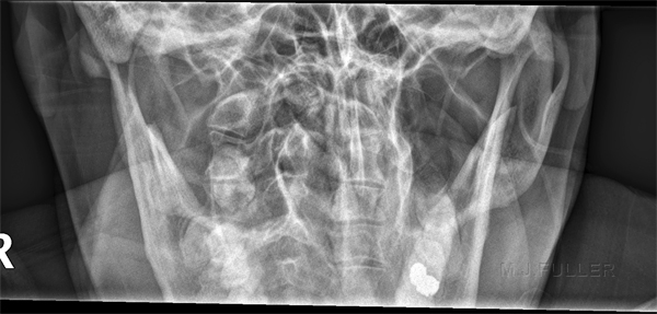 | It was decided that a slit Townes technique might demonstrate the right zygoma and would also have the benefit of a comparison view of the left zygoma. The slit Townes view demonstrated the left zygoma clearly but not the right. The imaging so far was increasingly suggestive of a depressed right zygoma fracture. The radiographer considered that a teacup view with further neck lateral flexion/head tilt should demonstrate the depressed fracture. |
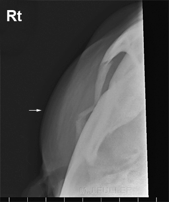 | The repeat teacup view demonstrated the depressed fracture of the patient's zygoma. The lateral flexion of the patient's neck is at the limit- any further lateral flexion of the patient's neck would cause the patients mandible to be projected clear of the outer table of the skull thereby overlapping the patient's zygomatic arch. This has occurred in this view but without detracting significantly from the diagnostic value of the image.
The radiographer considered that the patient might be referred for CT imaging of the facial bones. The radiographer consulted with the ED consultant and the plastic surgery registrar and it was decided that the TMJ views would not be performed in favour of CT imaging.
CT imaging uncovered a more extensive fracture than was revealed on plain film imaging. The fracture was seen to extend to the TMJ on the right. |
Comment
This case demonstrates a good grasp by the radiographer of a clinical approach to radiography. The radiographer's suspicion that the patient had sustained a depressed fracture of the right zygomatic arch was based on clinical considerations (patient history, clinical signs) as much as it was based on plain film evidence from the early imaging. The decision to perform a teacup view rather than a slit basal view of the right zygoma was a simple common-sense consideration. The decision to perform a second teacup view was also based on the notion that no matter how depressed the zygomatic fracture was, it could still be demonstrated on a teacup view.
Case 5
This patient presented to the Emergency Department after receiving multiple blows to the face during a fight. A selection of pre-operation and post-operation images are shown.
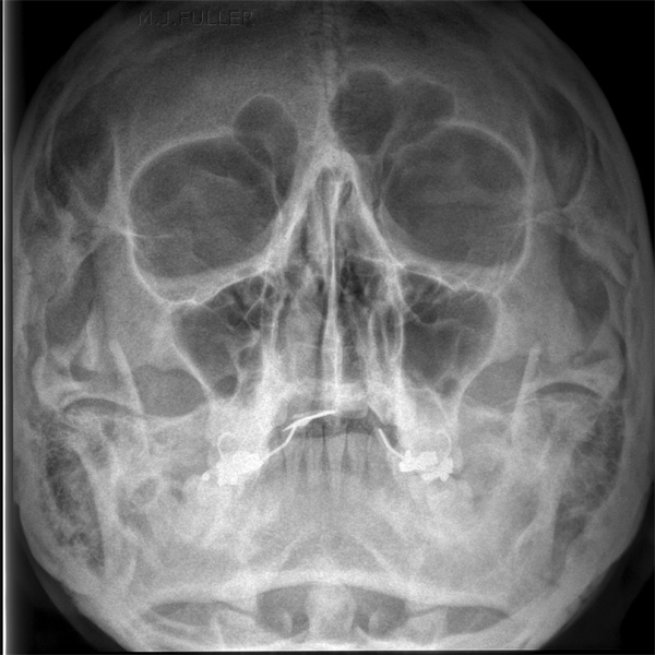 | The OM view image demonstrates no obvious fracture
C.R. image pre surgery |
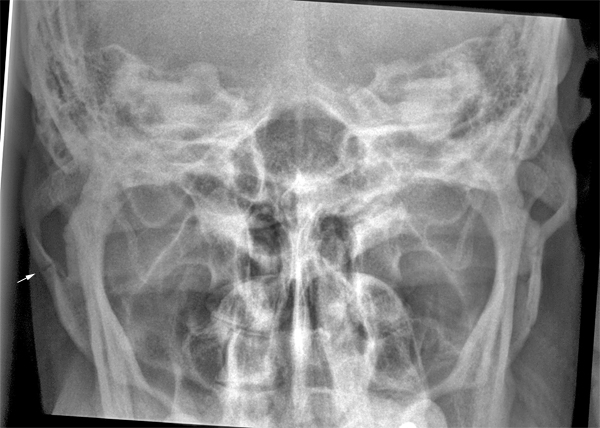 | The Slit Townes view suggests fractures of the right zygomatic arch.
C.R. image pre surgery |
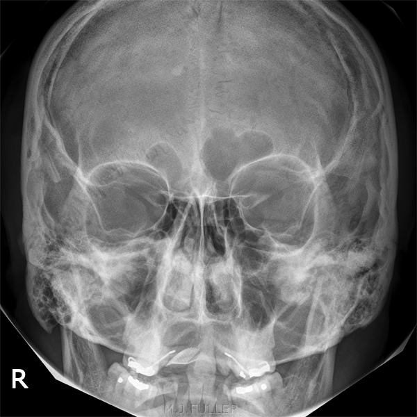 | The patient underwent a zygoma elevation operation on the right. This is the PA facial bones view image post operation.
D.R. image post-surgery |
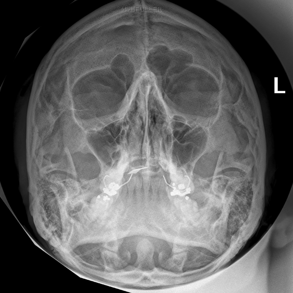 | Post op OM view of facial bones
D.R. image post-surgery |
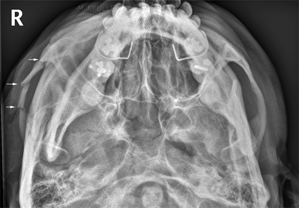 | This is the slit basal view of the zygomatic arches post zygoma elevation on the right. Note the effectiveness in demonstrating the zygomatic arches compared with the slit Townes view preop above.
D.R. image post-surgery |
DiscussionThe slit basal and teacup view techniques of the zygoma are learnable and reliable techniques. Once the positioning technique is mastered, the results are usually superior than that achieved with a slit Townes technique.
... back to the Applied Radiography home page
