Satisfaction Syndrome
Presentation
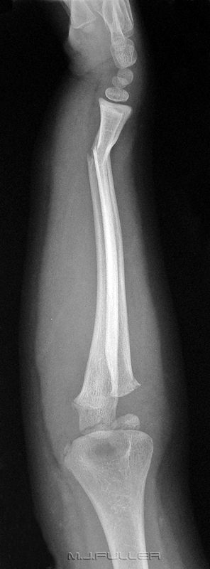
This girl has presented to the Emergency Department following an unwitnessed fall from her pushbike. She has an obvious deformity of her right wrist and forearm swelling and abrasions. Following a clinical examination the patient was referred for an X-ray examination of her right forearm. What abnormal finding(s) are revealed?
Imaging
Findings
The radiographer has performed ‘mixed’ views of the right forearm. That is to say, each view is a mixed AP and lateral projection. Whilst this is not ideal, it is widely considered as acceptable practice, particularly in paediatric radiography. From a pragmatic viewpoint, you often have no choice- this child was simply not prepared/able to pronate/supinate her hand.
Image 1.
It is clear that the patient has fractures of the distal radius and ulna with approximately 30 degrees of dorsal angulation. A fracture of the distal radius with dorsal angulation is commonly referred to as a Colle’s fracture. The danger at this point is that the radiographer/radiologist/clinician can experience a sense of satisfaction at successfully demonstrating the pathology that explains the patient’s symptoms. The motivation to undertake a detailed examination of the remainder of the demonstrated radiographic anatomy is potentially reduced. This is known as satisfaction syndrome or positive finding syndrome and has been the downfall of many a good X-ray examination.
On examination of the remainder of the anatomy, a fracture of the radial head is seen (Image 1, label 2). There is also a suggestion of a fracture of the coronoid(Image1, label 3) although this is not entirely convincing. There is a clearly visible fracture of the olecranon(Image 1, label 4). Are the structures labelled 5 and 6 (image1) fractures or normal developing epiphysis/apophysis? It is helpful to know the age of the patient and the normal development patterns of the elbow ossification centres. A commonly used mnemonic for remembering the elbow ossification centres is as followsNormal Elbow Ossification
C
Capitellum
R
Radial head
I
Internal (medial epicondyle)
T
Trochlea
O
Olecranon
L
Lateral Epicondyle
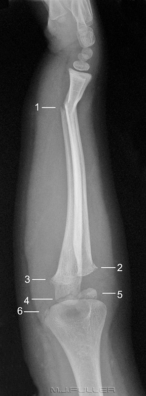
The ossification centre labelled 5 is the capitellum and appears to be normal. The next ossification centre to ossify is the radial head but this is not visible. The third ossification centre is that of the medial epicondyle (6) and it also appears to be normal. The medial epicondyle ossification centre has appeared before that of the radial head, out of the expected order. This is a relatively common occurrence. Despite this fact, using the CRITOL rule affords some confidence in determining that structures 5 and 6 are not avulsion fractures.Image 2There are a few irregular lucencies between the distal radius and ulna and these are almost certainly tracts of air. This indicates that the Colle’s fracture is a compound fracture - an important finding in terms of treatment. The compound nature of a fracture is not always obvious on clinical examination- the skin puncture can be subtle. There is also evidence of air in the soft tissues of the forearm. An olecranon fracture is clearly seen(3). There is also a second more subtle cortical breach of the olecranon articular surface. This may represent an additional fracture or may represent an element of comminution associated with the fracture labelled 3. The likelihood of a comminuted fracture of the olecranon is supported by the cortical double contour of the olecranon seen at the dorsal aspect of the olecranan (not marked).
It is noteworthy that the patient has an elbow effusion with anterior and posterior fat pad signs (labelled 4 and 5 respectively). This is an indicator of intra-articular injury although not a conclusive indicator of intra-articular bony injury. This finding is not a particularly helpful sign in the context of such an extensive elbow injury but should always be considered when assessing the lateral elbow image.Conclusion
There is a compound Colle’s fracture of the right wrist with approximately 30 degrees of dorsal angulation. There is also a radial neck fracture and at least one olecranon fracture which is likely to be comminuted.
....back to the applied radiography home page here


