Right Middle Lobe Collapse
Jump to navigation
Jump to search
False RML Collapse/Consolidation in Patients with Pectus Excavatum
False RML Collapse/Consolidation in Patients with Pericardial fat
Hidden RML Collapse/Consolidation
... back to the Applied Radiography home page
Introduction
Plain Film Appearance
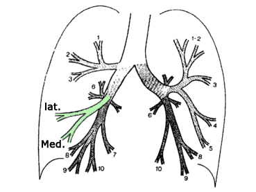
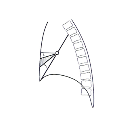
The Right Middle Lobe AnatomyThe right middle lobe (RML) is the odd one out- there is no 'left middle lobe' as such. The lingula segments of the left upper lobe are the equivalent of the RML. This is worth bearing in mind as the plain film pathological appearances of these lobes can demonstrate similar patterns..
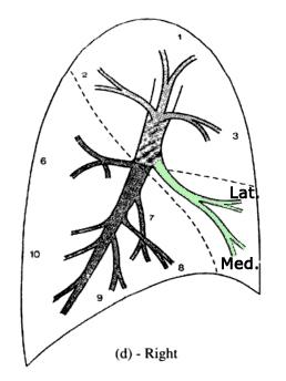
adapted from <a class="external" href="http://books.google.com.au/books?id=Bif0zpmEWtAC" rel="nofollow" target="_blank">By Fred W. Wright Radiology of the Chest and Related Conditions: Together with an Extensive Illustrative Collection of Radiographs CRC Press, 2002</a>The right middle lobe has two pulmonary segments which are situated side by side; the more lateral segment, approximates the size of its adjacent neighbour (medial segment). The medial segment abuts the right heart border medially , while the lateral segment extends to and comprises a portion of the lateral border of the right lung. <a class="external" href="http://lib.cpums.edu.cn/jiepou/tupu/atlas/www.vh.org/adult/provider/radiology/LungAnatomy/RightLung/RtLungSegAnat.html" rel="nofollow" target="_blank">http://lib.cpums.edu.cn/jiepou/tupu/atlas/www.vh.org/adult/provider/radiology/LungAnatomy/RightLung/RtLungSegAnat.html</a>.
When viewing chest radiographs with pathology involving the right middle lobe, it is important to think about the shape and position of the RML in three dimensions. This may not be easy at first. Note the description of the lobes is very approximate.
adapted from <a class="external" href="http://books.google.com.au/books?id=Bif0zpmEWtAC" rel="nofollow" target="_blank">By Fred W. Wright Radiology of the Chest and Related Conditions: Together with an Extensive Illustrative Collection of Radiographs CRC Press, 2002</a>
Important Characteristics of all Lobar Collapse
1. Collapse and consolation can occur independently or together2. Collapse can be partial or complete3. It is often not clear to what extent the appearance is due to collapse or consolidation or both. The degrees of each are often unclear.4. If a lobe is only partially collapsed and there is no accompanying consolidation, there may be no increase in opacity5. In cases of pure collapse, only when the collapse is virtually complete will there be a significant increase in density of the affected lung
Plain Film Appearance
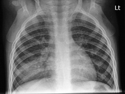
- The plain film appearance of collapse of the right middle lobe is often characterised by a loss of volume, an increase in density, and a loss of clarity of the right heart border.
- A partial RML collapse can be difficult to appreciate because the increase in opacity of the lobe can be superimposed over the heart shadow
A collapse of the lingula segment of the left upper lobe (LUL)
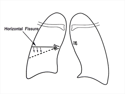
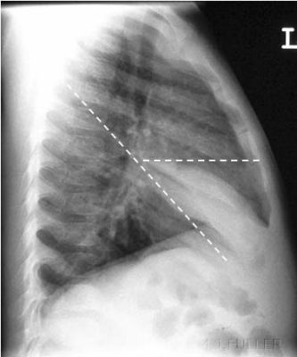
an appreciation of the collapse of the RML can be gained by consideration of the normal position of the horizontal and oblique fissure
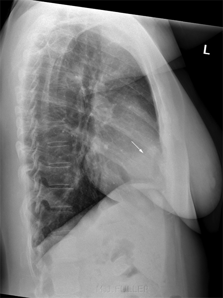
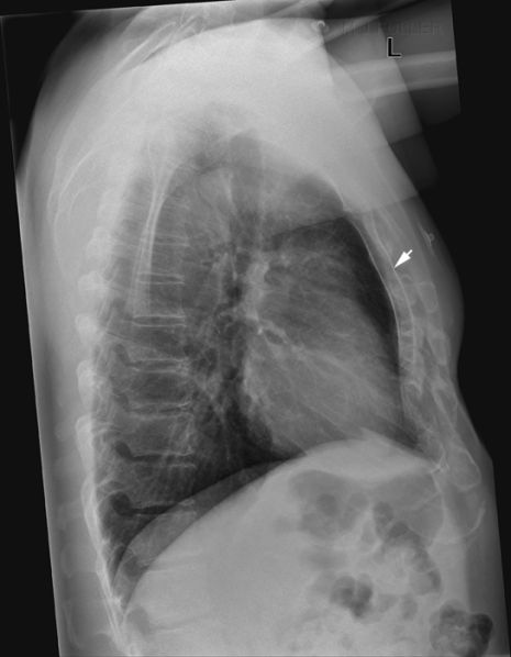
False RML Collapse/Consolidation in Patients with Pectus Excavatum
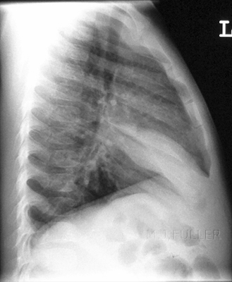
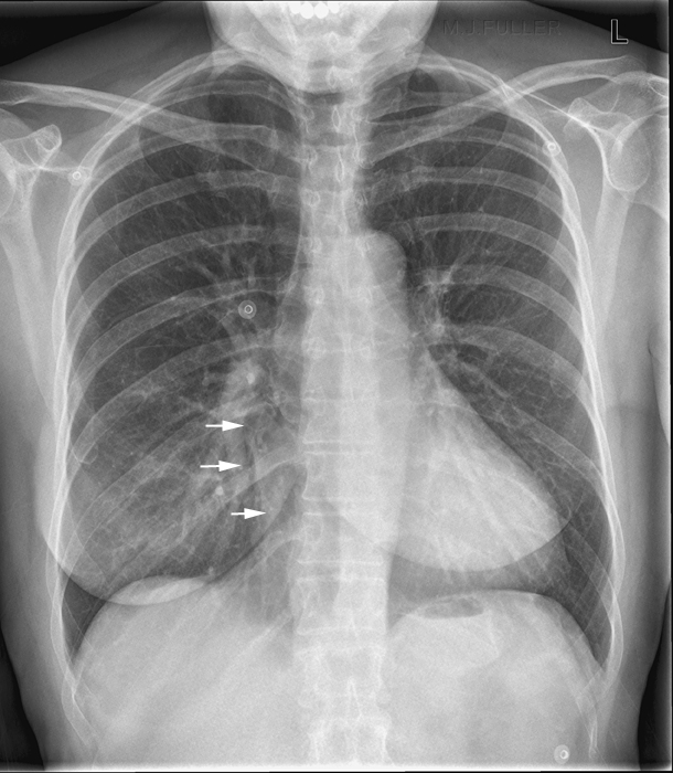
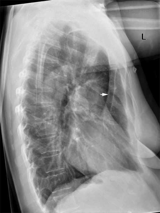
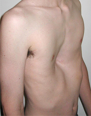
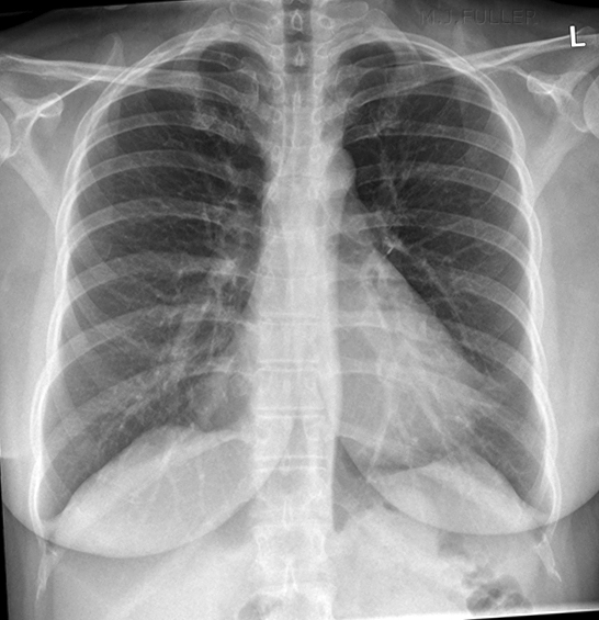
This patient presented for chest radiography with a history of recent facial nerve palsy. There is loss of visualisation of the right cardiac border. The lateral chest image demonstrates no abnormal RML opacity. There is evidence of pectus excavatum (depression of the sternum). Pectus excavatum is a known cause of false RML disease and is likely to be a contributing cause of the pseudo-silhouette sign on the PA chest image. In patients, with pectus excavatum the heart tends to be displaced towards the left as a result of the limited space between the depressed sternum and the spine.Other contributing factors could be pericardial fat (although none is seen in this patient) and overlying pulmonary vessel. Pectus Excavatum
<a class="external" href="http://en.wikipedia.org/wiki/File:Pectus1.jpg" rel="nofollow" target="_blank">http://en.wikipedia.org/wiki/File:Pectus1.jpg</a>
False RML Collapse/Consolidation in Patients with Pericardial fat
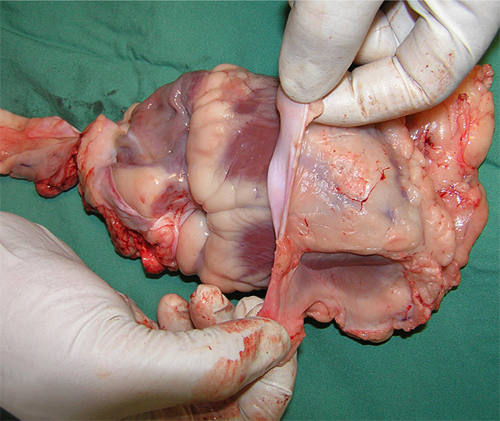
Pericardial Fat
<a class="external" href="http://jcmr-online.com/content/11/1/15" rel="nofollow" target="_blank">Validation of cardiovascular magnetic resonance assessment of pericardial adipose tissue volume</a><a class="external" href="http://jcmr-online.com/content/11/1/15" rel="nofollow" target="_blank">Adam J Nelson , Matthew I Worthley , Peter J Psaltis , Angelo Carbone , Benjamin K Dundon , Rae F Duncan , Cynthia Piantadosi , Dennis H Lau , Prashanthan Sanders , Gary A Wittert and Stephen G Worthley</a><a class="external" href="http://jcmr-online.com/content/11/1/15" rel="nofollow" target="_blank">Cardiovascular Research Centre, Royal Adelaide Hospital & Disciplines of Medicine and Physiology, University of Adelaide, Adelaide, SA, Australia</a>This is a sheep's heart with the pericardium partly dissected off. Note the pericardial fat.
Felson (<a class="external" href="http://www.amazon.com/Chest-Roentgenology-Benjamin-Felson/dp/0721635911/ref=sr_1_2?ie=UTF8&s=books&qid=1252240078&sr=1-2" rel="nofollow" target="_blank">Chest Roentgenology, W.B. Saunders, 1973, p55</a>) notes that "For reasons that escape me, fat in the thorax is often impossible to differentiate from water density. Perhaps the adjacent pulmonary gas density has something to do with this illusion." This is an interesting point given the clear differentiaton between fat and water density structures seenin the abdominal plain film.
Case 1
Hidden RML Collapse/Consolidation
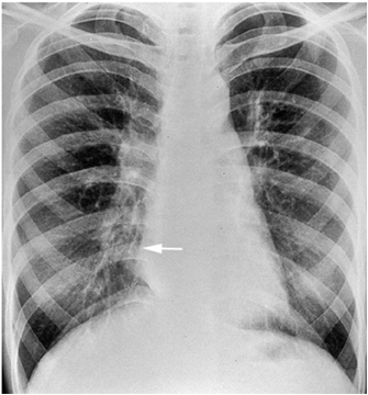
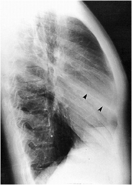
There is suggestion of some loss of visualisation of the right heart border
<a class="external" href="http://bjr.birjournals.org/cgi/content/abstract/74/877/89" rel="nofollow" target="_blank">K Ashizawa, MD, K Hayashi, MD, N Aso, MD and K Minami, MD
Lobar atelectasis: diagnostic pitfalls on chest radiography
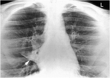
British Journal of Radiology 74 (2001),89-97, 2001</a>The arrowed structure looks like a fissure but it is not in the correct position for either the horizontal fissure nor the oblique fissure.
<a class="external" href="http://bjr.birjournals.org/cgi/content/abstract/74/877/89" rel="nofollow" target="_blank">K Ashizawa, MD, K Hayashi, MD, N Aso, MD and K Minami, MD
Lobar atelectasis: diagnostic pitfalls on chest radiography
British Journal of Radiology 74 (2001),89-97, 2001</a>The lordotic view demonstrates a completely collapsed RML (arrowed)
<a class="external" href="http://bjr.birjournals.org/cgi/content/abstract/74/877/89" rel="nofollow" target="_blank">K Ashizawa, MD, K Hayashi, MD, N Aso, MD and K Minami, MD
Lobar atelectasis: diagnostic pitfalls on chest radiography
British Journal of Radiology 74 (2001),89-97, 2001</a>
note- this is unlikely to represent an acute collapse
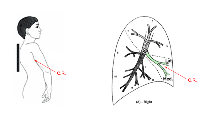
The centray ray is aligned to the orientation of the RML in the lordotic position. The lordotic position can reveal RML pathology which is hidden on the conventional PA and lateral views
... back to the Applied Radiography home page
