Radiography of the Bicipital Groove
Dedicated radiographic views of the bicipital groove of the humerus have lost popularity/utility over recent years. Despite the displacement of the view by other imaging modalities, bicipital groove can be a useful view in specific circumstances.
Anatomy
The biceps muscle has two muscle bellies, each one of them terminating in a corresponding tendon. The short bicipital tendon inserts into the coracoid process. The long bicipital tendon(LBT) inserts into the supraglenoid tubercle and/or the superior aspect of the glenoid labrum after arching over the humeral head. This tendon enters the joint capsule through the bicipital groove, formed between the lesser and greater humeral tuberosities. The LBT is maintained within the bicipital groove by the transverse humeral ligament. <a class="external" href="http://books.google.com.au/books?id=bHkNOvwbmocC&printsec=frontcover" rel="nofollow" target="_blank">Michael B. Zlatkin MRI of the shoulder, 2nd Ed. 2002</a>
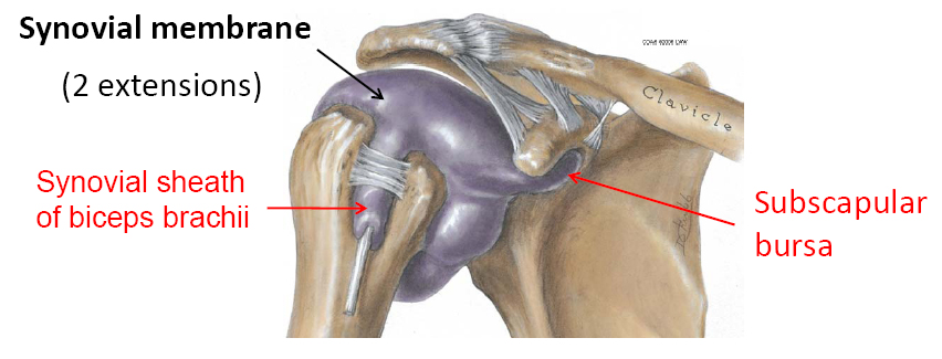
This graphic provides a good sense of the position of the bicipital groove Clinical examination for bicipital tendonitis.
The synovial membrane of the shoulder has two extensions- one covering the biceps brachii and the other inferior to the coracoid.
Radiography
<a class="external" href="http://books.google.com.au/books?id=ssX25YIdAFIC&printsec=frontcover" rel="nofollow" target="_blank">By Joseph P. Iannotti, Gerald R. Williams</a>
<a class="external" href="http://books.google.com.au/books?id=ssX25YIdAFIC&printsec=frontcover" rel="nofollow" target="_blank">Edition: 2, illustrated</a>
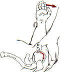
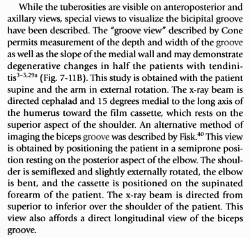
<a class="external" href="http://books.google.com.au/books?id=ssX25YIdAFIC&printsec=frontcover" rel="nofollow" target="_blank">Published by Lippincott Williams & Wilkins, 2006</a>This extract provides a description of the positioning for bicipital groove radiography.
<a class="external" href="http://www.ajronline.org/cgi/reprint/141/4/781" rel="nofollow" target="_blank">Cone et al</a> recommended the followingThis projection requires a supine position and a medial beam angulation of 15 to 25 degrees. Although we do not recommend that this film of the bicipital groove be included in every radiographic series of the shoulder, it may prove to be a useful adjunct in the assessment of some patients with bicipital tendon abnormalities as
well as those who have had shoulder trauma.
Research
The Bicipital Groove: Radiographic, Anatomic, and Pathologic Study
<a class="external" href="http://www.ajronline.org/cgi/reprint/141/4/781" rel="nofollow" target="_blank">Cone et al AJR:141, October 1983</a>
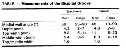
</a>Cone et al (1983) reported considerable variation in the bony dimensions of the bicipital groove.
Ultrasound vs Plain Film Radiograph of the Bicipital Groove
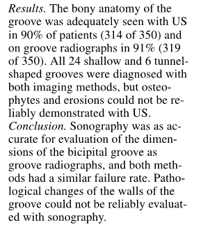
<a class="external" href="http://resources.metapress.com/pdf-preview.axd?code=64tu5rddvmyjchln&size=largest" rel="nofollow" target="_blank">Pekka U. Farin and Heikki Jeroma, The Bicipital Groove of the Humerus: Sonographic and Radiographic Correlation. Skeletal Radiology 1996. 25: 215:219</a> The bottom line appears to be that Ultrasound imaging is not good for demonstrating bony changes of the bicipital groove. It could be argued that Ultrasound and plain film imaging have complementary value with respect to imaging of the bicipital groove.
Pathology
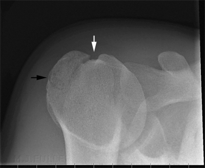
Several authors have noted that subluxations and dislocations of the bicipital tendon are more common in the presence of a shallow intertubenculan sulcus. <a class="external" href="http://www.ajronline.org/cgi/reprint/141/4/781" rel="nofollow" target="_blank">Cone et al AJR:141, October 1983</a>. Cone et al also noted that "...Bony spurs involving the bicipital groove seem to be relatively common."
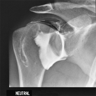
Case 1
Comment
Bicipital groove radiography is rarely performed and for good reason. The value of the view has been questioned by various studies. Despite these limitations, it is useful for radiographers to be aware of the view for its occasional utility.
Further Information
Fisk, C. (1965).Adaption of the technique for radiography of the bicipital groove, Radiol. Technol. 37:47-50.
... back to the Applied Radiography home page
