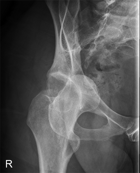
Radiography of Hip Dislocations

From wikiRadiography
Jump to navigation Jump to search
Introduction

Hip dislocations are commonly seen in patients with hip prosthesis and in cases of severe trauma. This page considers all aspects of radiography of hip dislocations


Case 1
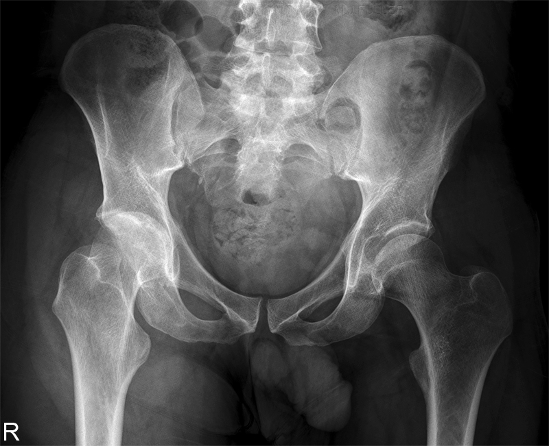
AP PELVISThis 53 year old male presented to the Emergency Department with right hip pain. His history was unknown. He was referred for hip radiography with a clinical diagnosis of right neck of femur fracture.

The right hip is in external rotation. The contour of the femoral head overlaps the acetabular rim suggesting hip dislocation.
RIGHT HIPThe radiographer proceeded to perform an LPO Judet view of the right hip.The hip dislocation was now clearly evident.
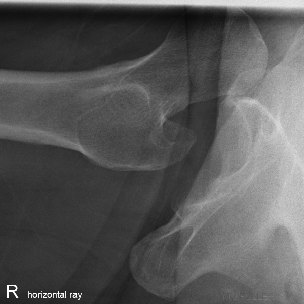
lateral H-ray right hipThe horizontal ray projection of the right hip demonstrates an anterior dislocation.