Proximal Tibiofibular Joint Dislocation

From wikiRadiography
Jump to navigation Jump to search
Introduction

Proximal tibiofibular joint dislocation is more commonly referred to as a fibular head dislocation. This injury is uncommon but is worthy of attention because it can be easily missed and because it can be mimicked by a combination of other pathology and projectional factors.


Case 1
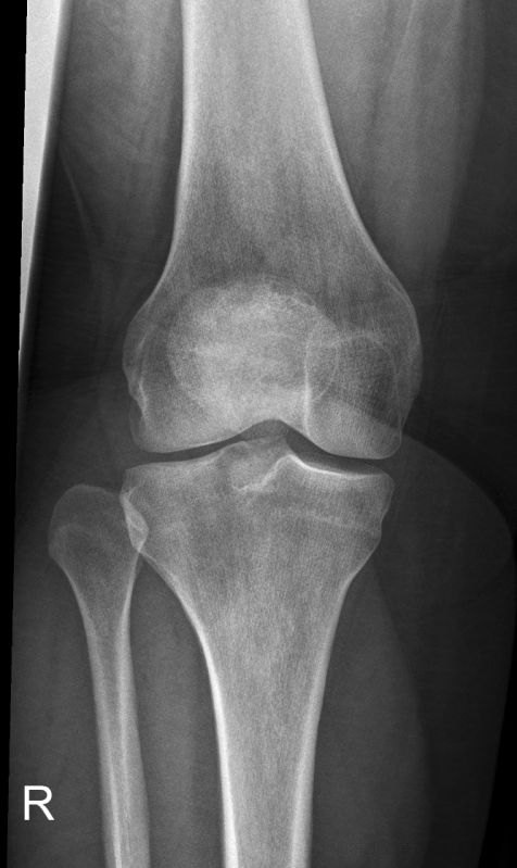
ap kneeThis patient presented to the Emergency Department following a right knee injury (details unknown). The radiographer noted lateral subluxation of the patella and compensated by internally rotating the knee thereby positioning the patellar centrally over the femoral condyles. In doing so, the fibular head has come into profile giving the appearance of a dislocated fibular head.
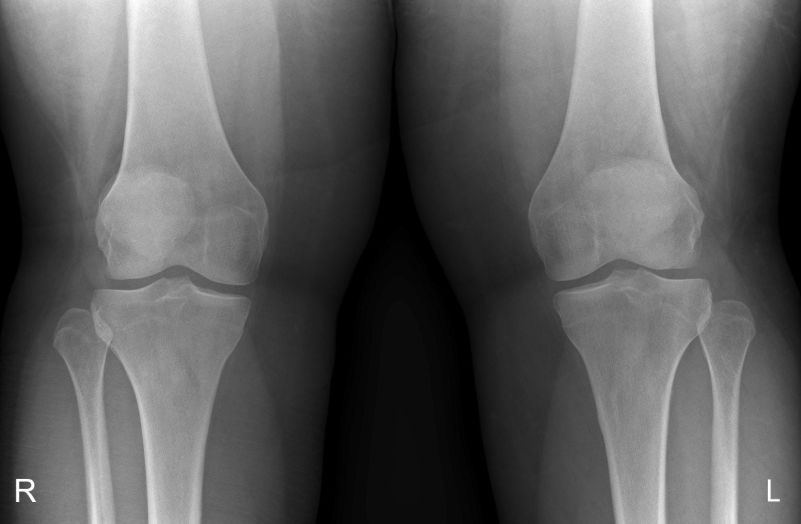
AP kneesThe radiographer was aware of the potential for misdiagnosis of a proximal tibiofibular joint dislocation. A single exposure AP projection of both knees was performed for the purposes of comparison. The right knee was positioned in a true AP position demonstrating the lateral subluxation of the right patella. The appearance of the proximal tibiofibular joints is comparable suggesting that the right proximal tibiofibular joint is normally aligned.
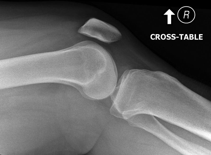
lateral kneeThe lateral knee projection image is unremarkable. There is a mall knee effusion.
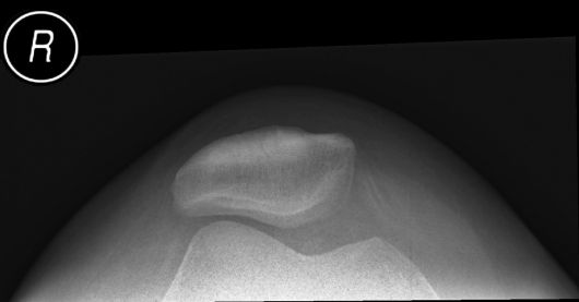
skyline kneeThe lateral subluxation of the patella is demonstrated on the skyline projection image.