Posterior Malleolus Fractures

From wikiRadiography
Jump to navigation Jump to search
Introduction

Radiographic detection of posterior malleolus fractures of the distal tibia can be difficult. Consideration of: the mechanism of injury; clinical presentation; radiographic soft tissue signs; pattern recognition; and positioning traps can assist in avoiding missed fractures.


Mechanism of Injury

"Posterior malleolus fractures may occur in isolation or, more commonly, in association with bimalleolar or trimalleolar fracture patterns. These fractures result from posterior tibiofibular ligament avulsion, or bony impaction from the talus." <a class="external" href="http://www5.aaos.org/oko/description.cfm?topic=TRA024&referringPage=mainmenu.cfm" rel="nofollow" target="_blank">(Orthopedic Knowledge Online, Treatment of Posterior Malleolus Fractures )</a>


Anatomy
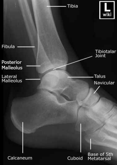
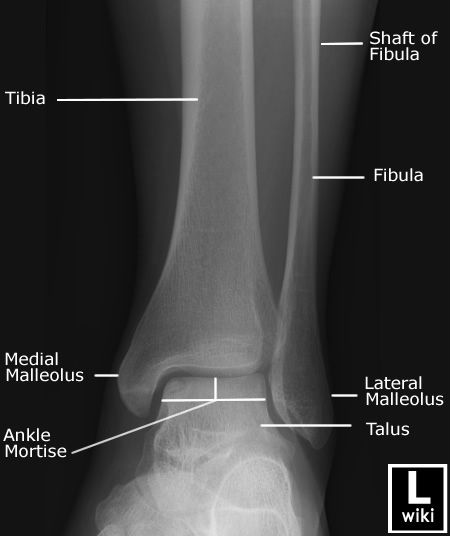
lateral ankle anatomyRadiographic Anatomy - Ankle Mortise


The Soft Tissue Signs

The ankle has three important potential soft tissue signs.

1. Soft Tissue Symmetry

ankle trauma 4 oblnormal ankle
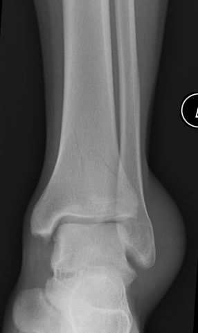
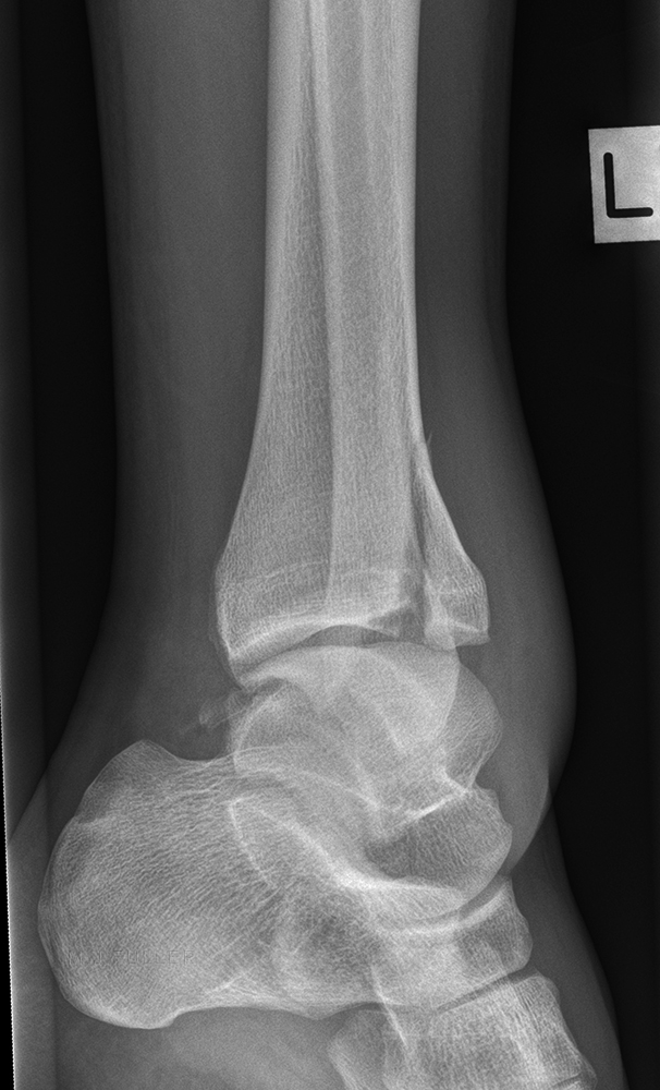
The thickness of the soft tissues overlying the medial malleolus and lateral malleolus should be approximately equal. This patient has increased soft tissue thickness over the lateral malleolus associated with a largely undisplaced spiral fracture of the distal fibulaCompare with this normal ankle


2. Ankle Effusion- The Teardrop Sign

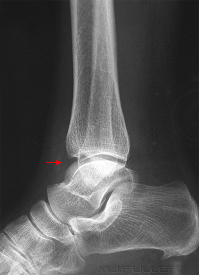
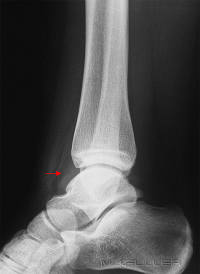
An ankle effusion suggests a significant injury to the ankle joint. The anterior and posterior juxta-capsular region of a normal ankle joint should appear as a fat-like density. In the presence of an ankle effusion, the capsule can become distended and may appear to have a more fluid-like density

Soft Tissue Signs in Orthopaedics - wikiRadiography

Soft Tissue Signs in Orthopaedics - wikiRadiography
normal ankle demonstrating a fat -like density (arrowed) abnormal ankle demonstrating a fluid-like density (arrowed)


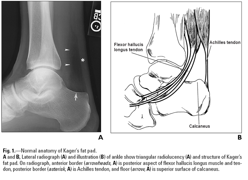
3. Kager's Fat Pad
Soft Tissue Signs in Orthopaedics - wikiRadiography

<a class="external" href="http://www.ajronline.org/cgi/content/full/182/1/147" rel="nofollow" target="_blank">Justin Q. Ly and Liem T. Bui-Mansfield Anatomy of and Abnormalities Associated with
Kager’s Fat Pad AJR:182, January 2004
</a>

It is not uncommon for ankle injuries to involve Kager's fat pad. A careful examination of the density, shape and borders of Kager's fat pad can provide indicators of bony injury to the ankle. An abnormal Kager's fat pad does not indicate definite bony injury to the ankle.


Misty Mountains Sign
ankleankle mountain peak signankle mountain peak sign
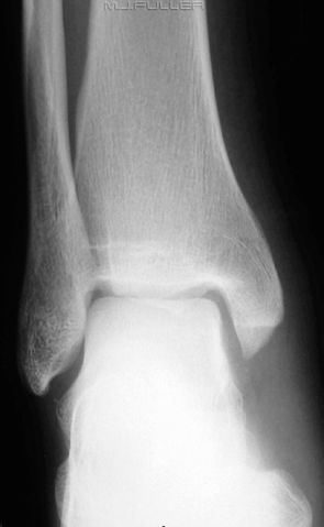
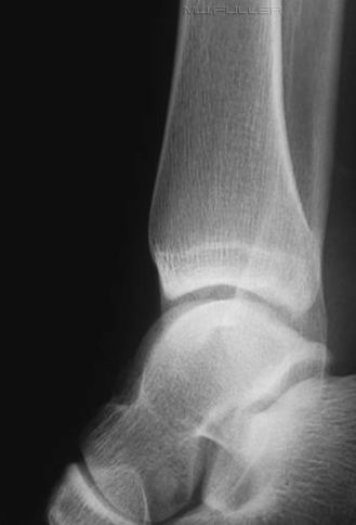
These 3 patients all have posterior malleolus fractures and misty mountains sign.
Mountain Peaks in a mist
mountain peak sign

adapted from <a class="external" href="http://z.about.com/d/taoism/1/0/2/1/-/-/JadeDragonMountain12.jpg" rel="nofollow" target="_blank">http://z.about.com/d/taoism/1/0/2/1/-/-/JadeDragonMountain12.jpg</a>
Coronal plane distal tibial metaphysis fractures have a plain film appearance which resembles mountain peaks faintly visible through mist. If this sign is not recognised , patients with tibial metaphysis fractures can have their fractures missed. Radiographers who are able to recognise this sign will be able to perform the necessary supplementary projection imaging required at the time of the acute presentation.


The Positioning Trap

A poorly positioned lateral ankle can either hide a posterior malleolus fracture or reveal a posterior malleolus fracture. The following case studies demonstrate both types of cases

Case 1
The Lateral Ankle Trap - wikiRadiographyThis patient presented to the Emergency Department with a painful and swollen right ankle. The mechanism of injury was unknown. The referring doctor requested a right ankle x-ray examination to rule out any bony injury.

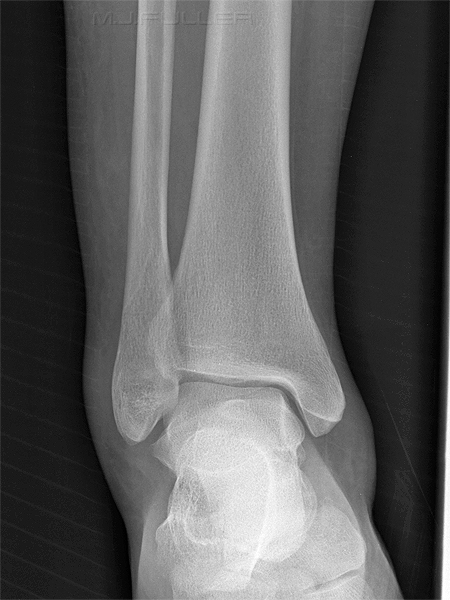
The mortise ankle projection image demonstrates no displaced fracture.
The Lateral Ankle Trap - wikiRadiographyThe lateral ankle projection image also demonstrates no displaced fracture.
The Lateral Ankle Trap - wikiRadiographyThe repeat lateral ankle has projected the distal fibula off the posterior malleolus of the distal tibia revealing a tibial posterior malleolus fracture.

Discussion
The over-rolled ankle positioning error is particularly risky in terms of posterior malleolus fractures. It would be reasonable to ask if this is simply a freak occurrence and unworthy of consideration over and above being quaint and quirky?


Case 2

The Lateral Ankle Trap - wikiRadiography
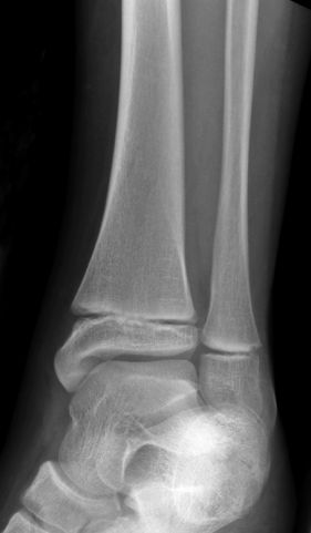
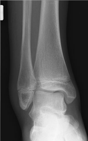
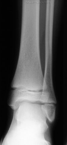
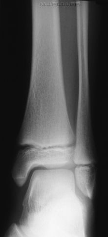
This child has presented to the Emergency Department following an unwitnessed fall. The patient was assessed and ankle X-ray imaging is requested

The AP ankle projection image demonstrates no displaced fracture
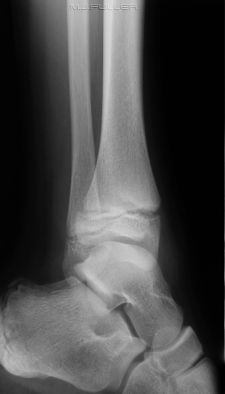
The Lateral Ankle Trap - wikiRadiographyThe oblique ankle projection image demonstrates no displaced fracture

The Lateral Ankle Trap - wikiRadiography

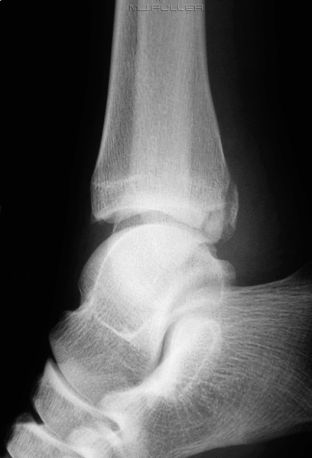
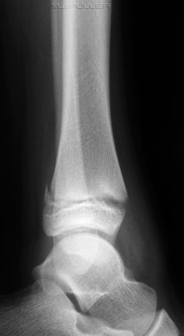
The lateral ankle projection image demonstrates no displaced fracture

Image Evaluation
The lateral ankle is over-rolled causing the distal fibula to be superimposed over the posterior malleolus. The radiographer considered this image worthy of repeating. The repeat image is shown below.
The Lateral Ankle Trap - wikiRadiography

This lateral ankle is in a good position and reveals a Salter-Harris II fracture of the posterior distal tibia. (? SH I also)


Case3
ap anklePatient History
Twisted right ankle. Not weight-bearing

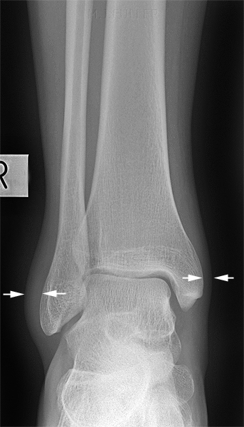
The AP ankle projection image demonstrates soft tissue thickening laterally. There is no evidence of an associated displaced fracture of the fibula


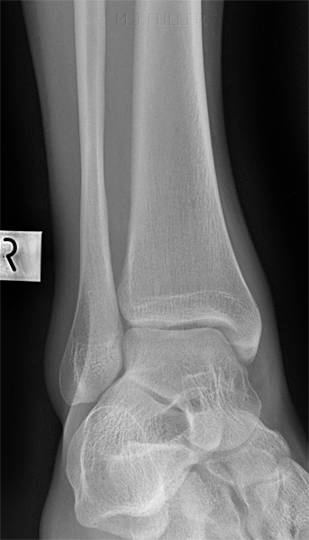
oblique ankleThe oblique ankle image demonstrates no displaced fractures
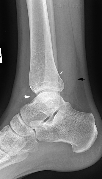
lateral ankleThe lateral ankle image demonstrates the following

  • ankle effusion (lower white arrow)
  • abnormal Kager's fatpad (black arrow)
  • very subtle defect in cortex of posterior malleolus of tibia (top white arrow)


LATERAL ANKLEDiscussion
The radiographer assessed the routine views in the context of the patient's mechanism of injury and clinical features. She considered there was a reasonable possibility that this patient had sustained a fracture of the posterior malleolus of the tibia and there was a fair chance of demonstrating such a fracture by performing off-lateral views of the ankle.

This is an under-rotated lateral. No displaced fracture is demonstrated

lateral ankle

bingo! the over-rotated lateral position demonstrates a minimally displaced fracture of the posterior malleolus. Interestingly, this a very similar position to that which failed to demonstrate the posterior malleolus in case 1 and case 2.

It is noteworthy that when the radiographer returned to the patient to perform these supplementary views, the referring doctor was discharging the patient with an ankle sprain. Following the demonstration of the fracture on the supplementary views, the patient had a plaster cast applied to his ankle and a follow-up appointment was made for an orthopaedic clinic review in 2 weeks.

If you think you can't make a difference to patient outcomes as a radiographer...I would beg to differ!


Case 4

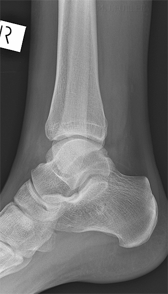
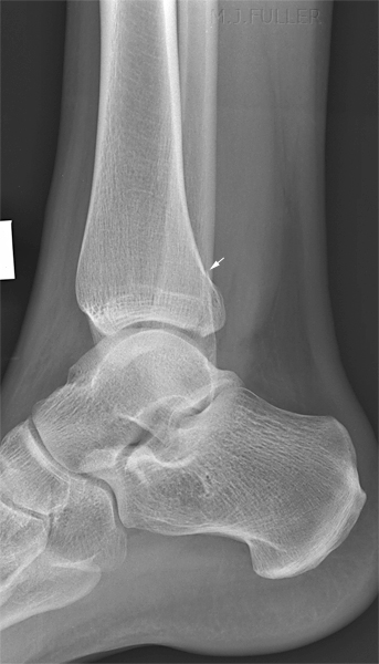
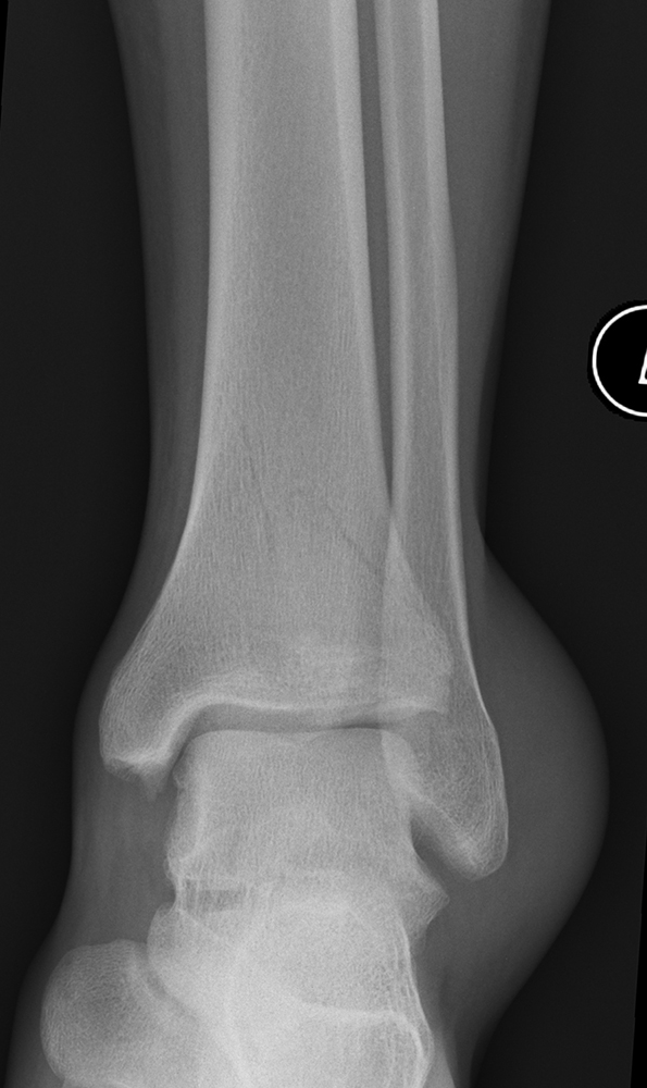
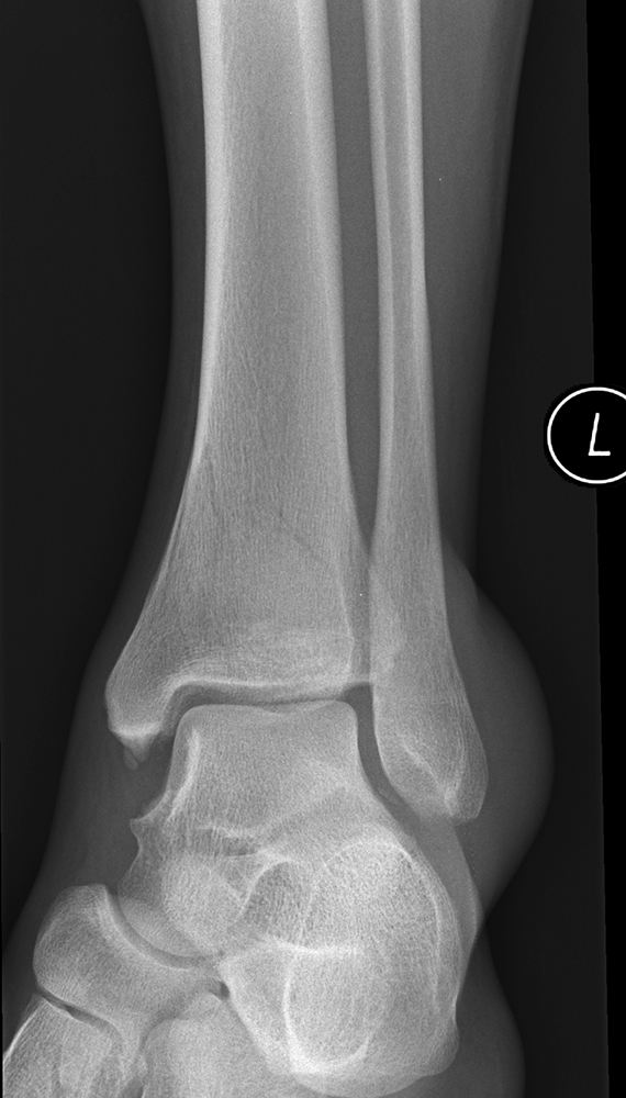
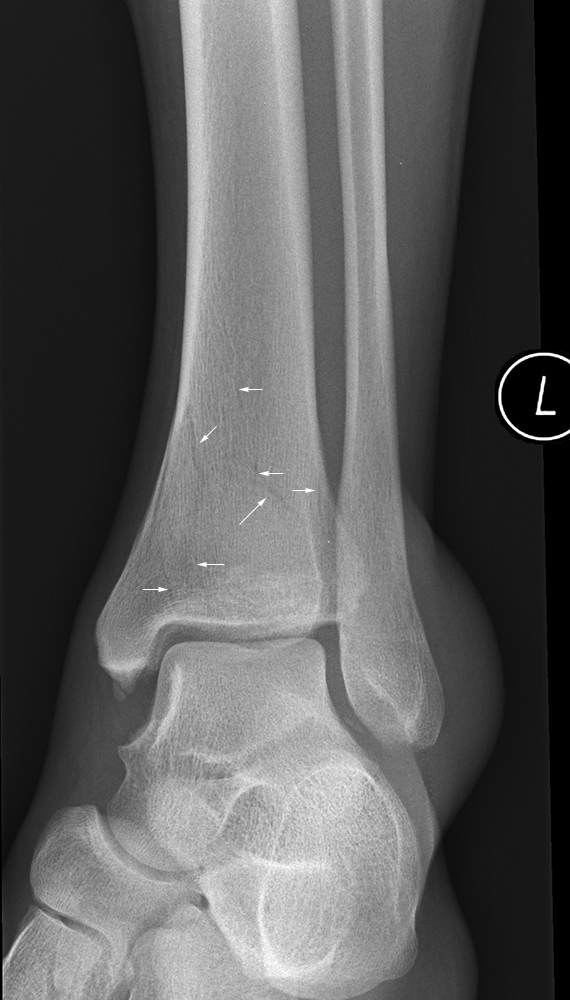
ankleThis 21 year old male presented to the Emergency Department following falling awkwardly onto his left ankle. On examination his ankle was found to be painful and there was swelling over the lateral malleolus. He was unable to weightbear on his left ankle. He was referred for left ankle radiography.
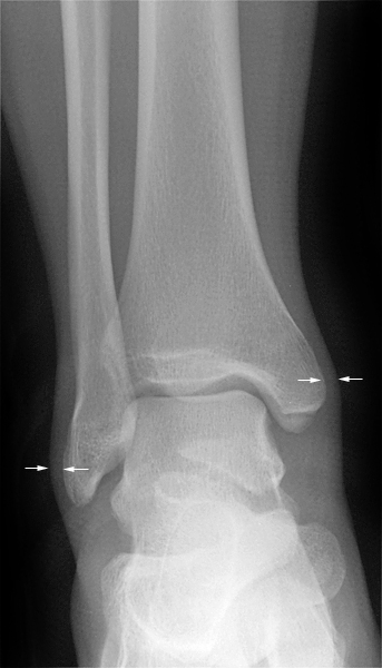
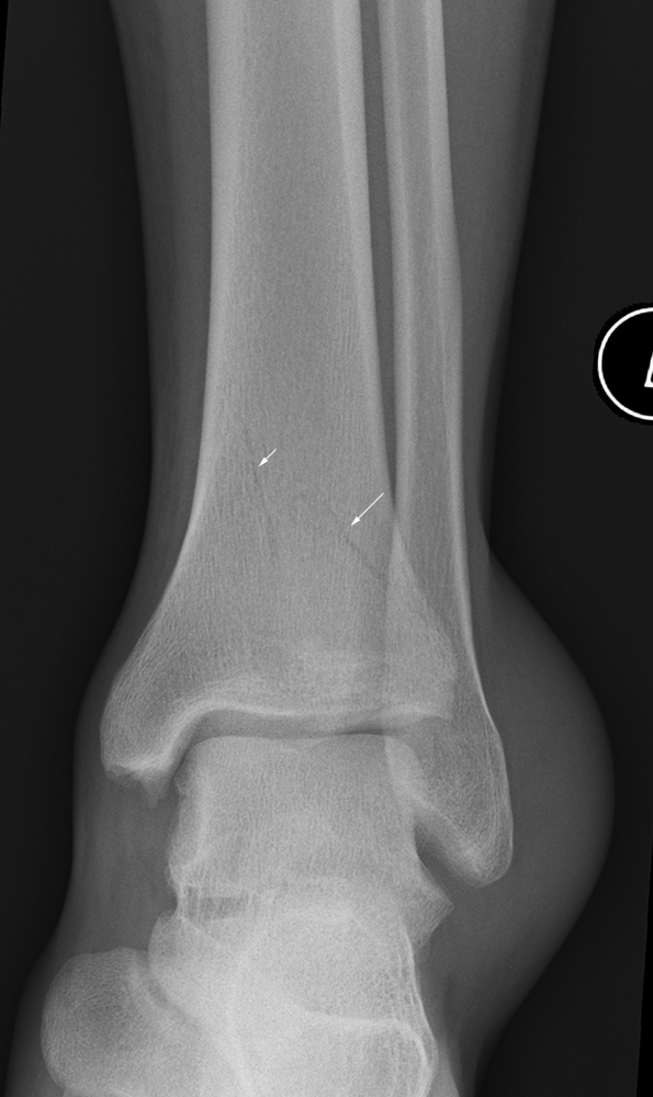
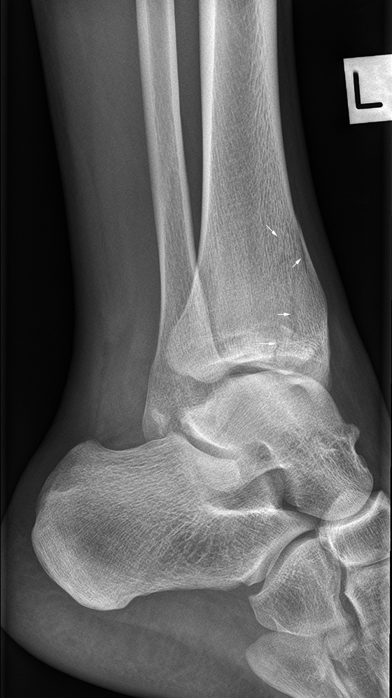
ankleThere is a subtle irregular lucency through the distal tibial metaphysis suggestive of a fracture in the coronal plane (mountain peak sign).
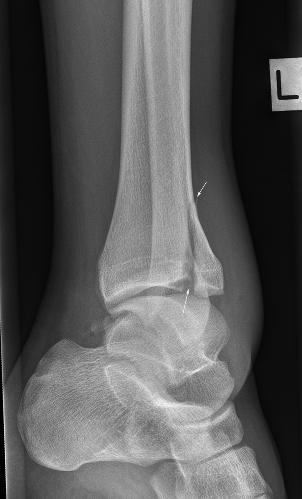
ankleThe oblique ankle projection image also demonstrated irregular lucent line suggesting a coronal plane fracture
ankleThe fracture lines (arrowed) indicate some complexity of the fracture, possibly associated with comminution.
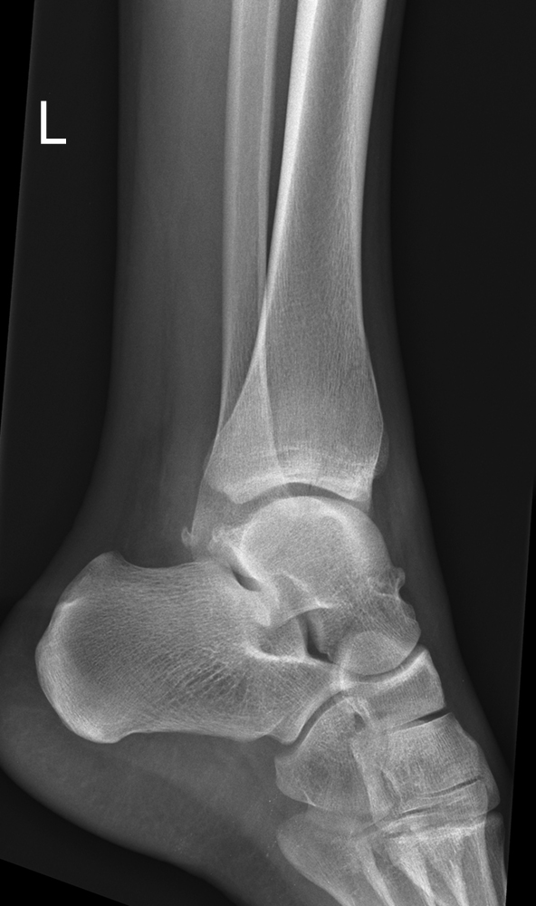
ankleThe lateral ankle projection image demonstrated subtle fracture lines suggesting that the fracture may be located anteriorly rather than the more common posterior malleolus fracture.
ankleOn discussion with the referring doctor, the management plan for this fracture was backslab and review at fracture clinic at a date to be determined. The radiographer considered that the imaging had not adequately demonstrated the fracture and that supplementary views may be helpful. The fracture line appeared to be orientated in a coronal plane. It was likely that off-lateral projections may align the X-ray beam with the plane of the fracture.

There appears to be a fracture of the posterior process of the talus.

This is essentially a deliberate malpositioned lateral ankle projection.
ankleThe fracture is again demonstrated but no additional information is provided
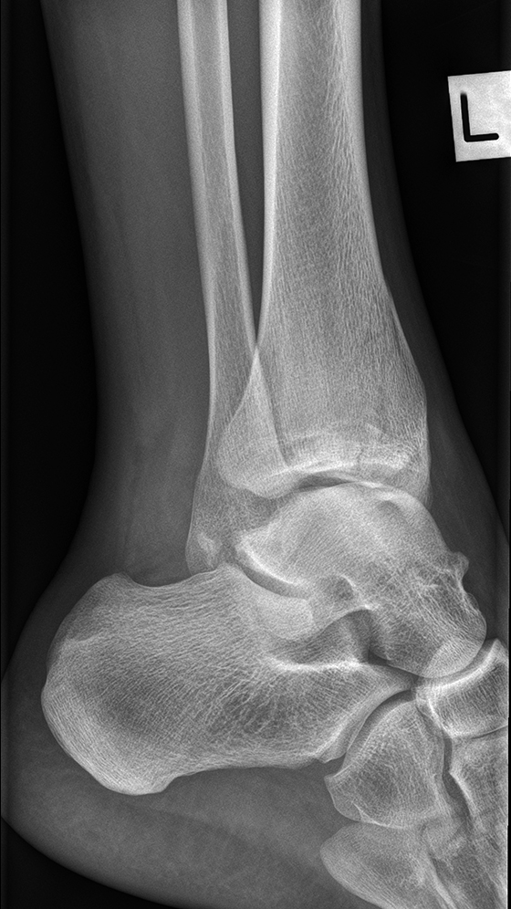
ankleThis is the opposite malpositioned lateral ankle projection.
ankleThis projection demonstrates an osteochondral fracture with significant displacement of the anterior fragment (arrowed). The patient received orthopaedic review in ED and was referred for a pre-operative planning CT.

Comment

The radiographer demonstrated considerable skill and judgement in performing supplementary views in a patient whose fracture had been demonstrated to the satisfaction of the referring doctor. It was within the radiographer's scope of practice to perform supplementary projections which proved to be pivotal in changing the patient's management from conservative to surgical. The mountain peak sign can be seen in any AP (ish) ankle projection of coronal plane distal tibial fractures... they are no always posterior malleolus fractures


Discussion


Posterior malleolus fractures of the distal tibia are easily missed, particularly when they exist in isolation and there is minimal displacement of the fragment. There is no guarantee that a perfectly positioned lateral ankle will demonstrate a patient's fracture. Indeed, a perfect lateral ankle position may hide the fracture. A consideration of the patient's mechanism of injury, clinical features, and radiographic soft tissue signs will decrease the likelihood of a missed fracture. It is noteworthy that case three was performed by a newly graduated radiographer. The level of proficiency demonstrated in this case is achievable by junior radiographic staff.


... back to the Applied Radiography home page