Pleural Effusions
Introduction
Pleural effusions demonstrated with chest radiography are nothing if not commonplace. For the radiographer there can be more to imaging a pleural effision than you might think. This page is dedicated to the radiographic appearances of pleural effusions, appearances that mimic pleural effusions and the radiographic techniques for demonstrating pleural effusions.
Definition of terms
A pleural effusion is a collection of fluid in the pleural space.
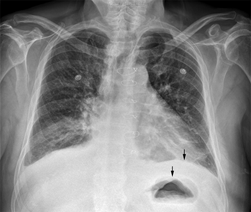
Pleural Effusion on Erect Chest Images
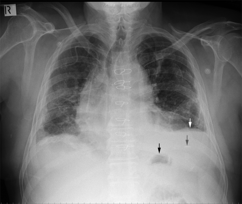
Pleural Effusion on Supine Chest Images
False Effusions
Subpulmonic Effusion
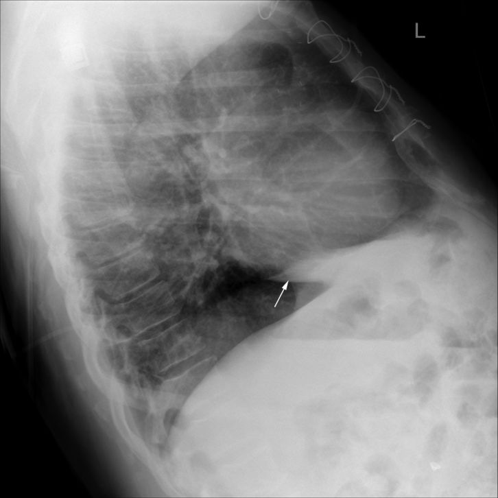
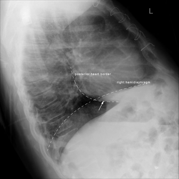
A subpulmonic effusion refers to a plural effusion which is loculated/contained between the diaphragm and the lung within the pleural space.
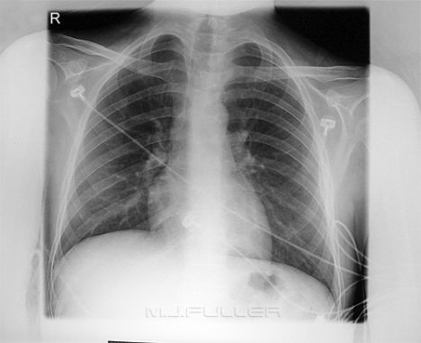
Subpulmonic effusion Normal Chest
Note the flat looking "hemidiaphragm", particularly medially. When you compare this appearance with the normal chest, the differences may appear subtle. The raised right hemidiaphragm, the flattened look of the diaphragm and the concave horizontal fissure suggest a possible subpulmonic effusion
<a class="external" href="http://www.med.yale.edu/intmed/cardio/imaging/cases/pleural_effusion_subpulmonic/index.html" rel="nofollow" target="_blank">http://www.med.yale.edu/intmed/cardio/imaging/cases/pleural_effusion_subpulmonic/index.html</a>
Compare this normal right hemidiaphragm shape to that of the patient with the right sided subpulmonic effusion.
A subpulmonic effusion can be mimicked by "subdiaphragmatic abscess, enlarged liver, paralysis and eventration of the diaphragm, and ascites
Case 1
| | |
|
| | |
|
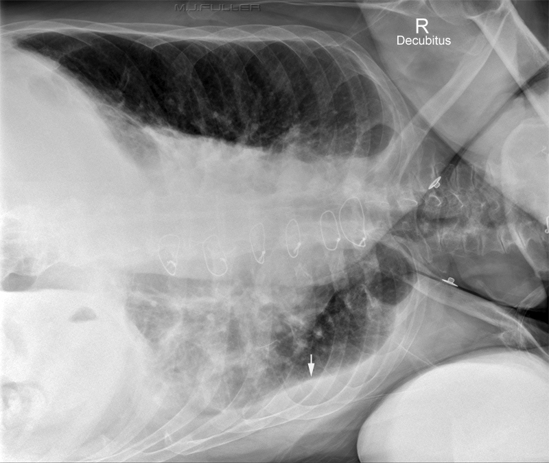
Decubitus Chest Technique for Pleural Effision
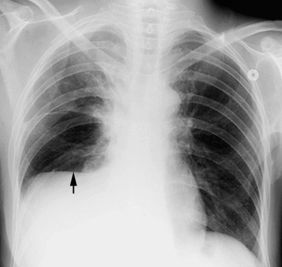
The left lateral decubitus chest image demonstrates fluid in the pleural space (arrow). Note that this is termed a left lateral decubitus view even though it is marked "R decubitus".
Artifacts and Normal Variants Simulating Pleural Effusion
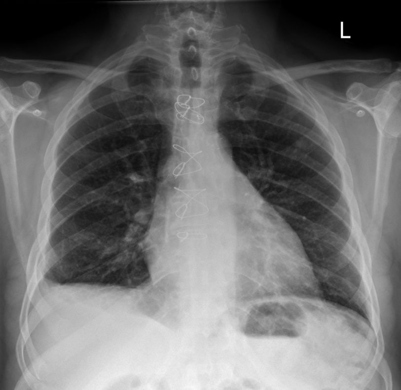
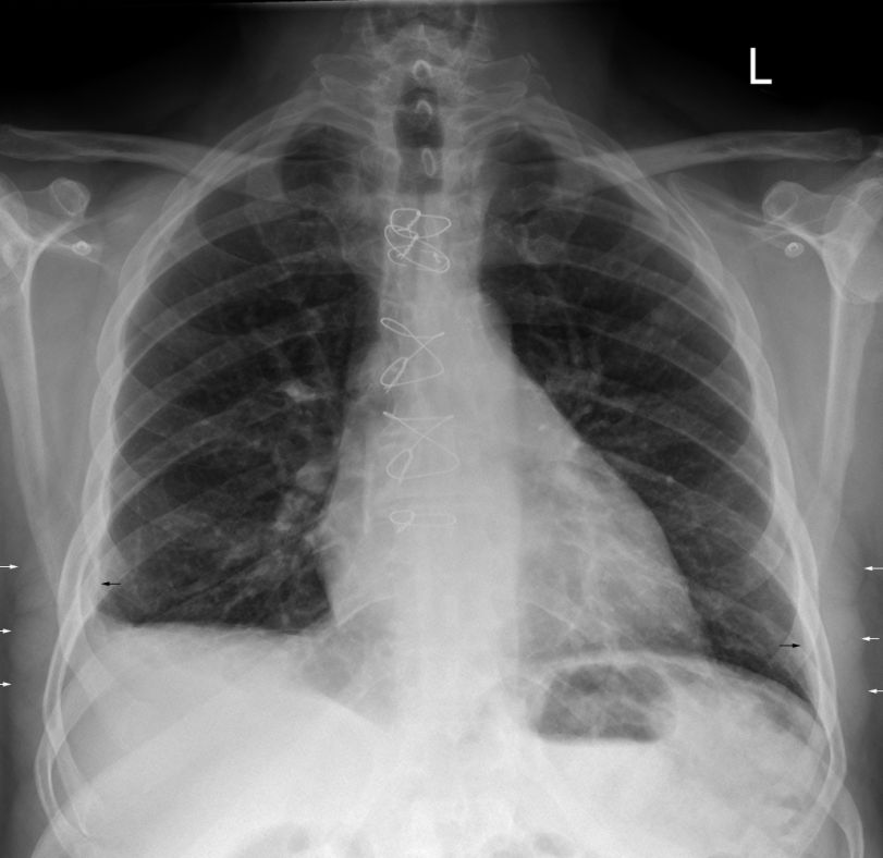
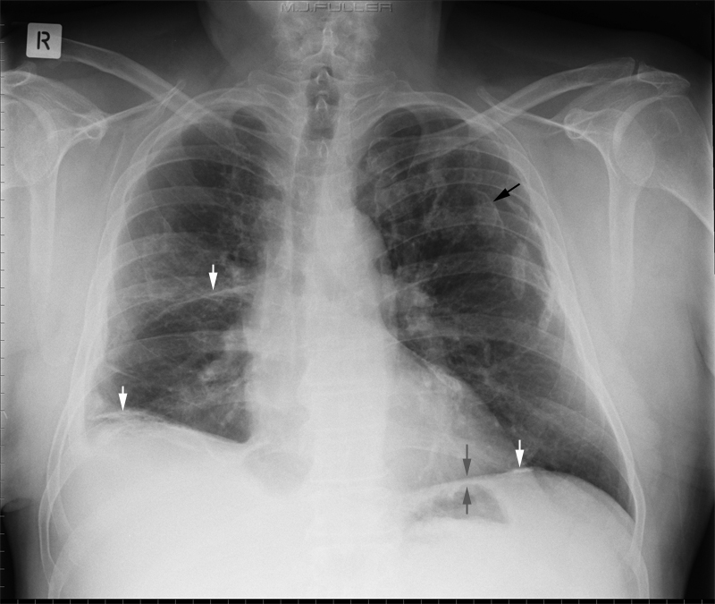
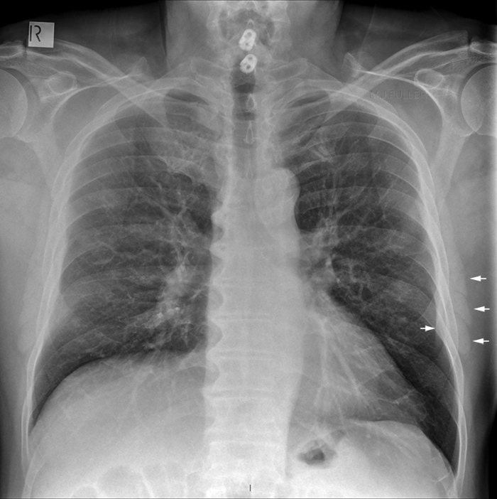
This patient has prominent serratus muscles bilaterally. This apparance can be mistaken for pleural effusion or pleural thickening (single white arrow).
The Serratus anterior muscle can produce an opacity that may resemble pleural tickening. This is sometimes referred to as a "bowling-pin" silhouette.
"When the muscle is well developed, the medial edge of this silhouette may be superimposed upon the air shadow of the lung in a variety of ways. When it overlies the apex of the lung, it gives rise to the companion shadow; when overlying the midlateral lung edge and costophrenic angle it may mimic pleural and/or extrapleural disease. Recognition of the various possible patterns is important to prevent overdiagnosis of disease, particularly asbestosis."
Gilmartin D.The serratus anterior muscle on chest radiographs. Radiology. 1979 Jun;131(3):629-35.<a class="external" href="http://en.academic.ru/pictures/enwiki/83/Serratus_anterior.png" rel="nofollow" target="_blank">http://en.academic.ru/pictures/enwiki/83/Serratus_anterior.png</a>
... back to the Wikiradiography home page
...back to the Applied Radiography home gae
