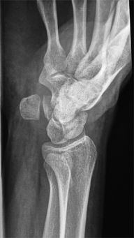
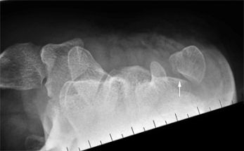
Pisiform Fractures
Introduction
Mechanism of InjuryThe pisiform is uncommonly fractured. When the radiographer is presented with a suspected pisiform fracture, a knowledge of the appropriate supplementary views will potentially assist in confirming the fracture.
The pisiform is typically injured by a fall on an outstretched hand in a dorsiflexed position, with the impact on the hypothenar eminence <a class="external" href="http://emedicine.medscape.com/article/828746-overview" rel="nofollow" target="_blank">Bryan C Hoynak, Laura Hopson, MD, e medicine</a>
Anatomy
The pisiform is a sesamoid bone within the tendon of the flexor carpi ulnaris. It articulates only with the triquetrum and lies near the deep ulnar nerve and artery.
Incidence
The average incidence of pisiform fractures is 0.2% of all carpal fractures and approximately half of them are isolated fractures.
<a class="external" href="http://www.ispub.com/ostia/index.php?xmlFilePath=journals/ijra/vol3n2/pisiform.xml" rel="nofollow" target="_blank">M. Tayfun Altınok MD. </a> <a class="external" href="http://www.ispub.com/ostia/index.php?xmlFilePath=journals/ijra/vol3n2/pisiform.xml" rel="nofollow" target="_blank">, Kadir Ertem MD , Ahmet Sığırcı MD , Alpay Alkan MD An Isolated Acute Pisiform Fracture: Usefulness Of MR Imaging
</a>
Radiography
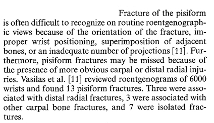 | ||
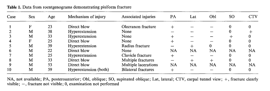
| <a class="external" href="http://resources.metapress.com/pdf-preview.axd?code=g308wk5172125n27&size=largest" rel="nofollow" target="_blank">Michael A Fleege, Pisiform Fractures, Skeletal Radiology, 1991: 20 p161</a> | ||
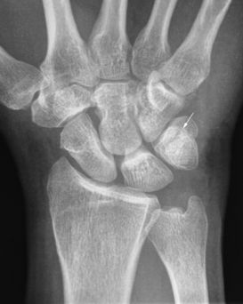
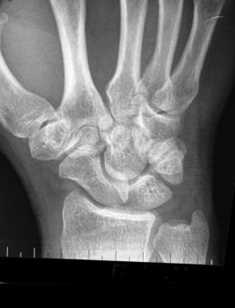
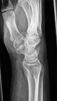
Demonstration of pisiform fractures will usually require dedicated radiographic techniques. In particular, a 20 degree supinated lateral wrist and carpal tunnel view may be helpful. In a study by Fleege et al (1991), the PA view was also found beneficial in demonstrating pisiform fractures.
<a class="external" href="http://resources.metapress.com/pdf-preview.axd?code=g308wk5172125n27&size=largest" rel="nofollow" target="_blank">Michael A Fleege, Pisiform Fractures, Skeletal Radiology, 1991: 20 p161</a>
...back to the Wikiradiography home page
...back to the Applied Radiography home page
