Physics of the X-Ray Tube
Jump to navigation
Jump to search
This page will deal with how the x-ray tube works and what happens inside the tube when you press buttons on the control console.
As I discovered last year, it is possible to hold a degree in medical imaging and still have no grasp on the fundamentals. The radiographer in question subsequently failed their PDY.
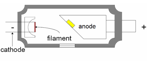
First things first, what does an x-ray tube look like? The first picture is a detailed picture of the inside of a tube. The second is a far more simplified version for teaching purposes.
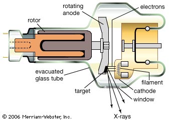
The x-ray tube is essentially a massive valve, or switch, completing an electric circuit. The x-ray tube consists of a cathode and an anode within a vacuum. The tube is enclosed within a housing. The negative end of the x-ray tube is called the cathode, and the positive end is called the anode. In any electric circuit, current flows from the negative side of a circuit to the positive side, and it is no different when an x-ray tube is involved.
The cathode is the negative side of the x-ray tube. The purpose of the cathode is to produce the thermionic cloud, conduct the high voltage current, and focus the emitted electrons (1). The cathode is more correctly called the cathode assembly. It consists of the filament (or filaments), the focusing cup and all the associated wiring. It is important to note that in radiography, the terms cathode and filament are generally interchangeable. Most radiographers, when talking about the cathode, are actually talking about the filament.
Discussion of the associated wiring is relatively questionable, except when discussing (a) focal spot size selection or (b) grid-biased (or grid-controlled or grid-pulsed) tubes. We will cover (a) later but not (b).
The anode assembly is the positive end of the x-ray tube. The anode assembly consists of the anode, the stator and the rotor. As a whole, the anode assembly is important as the source of the x-rays, as the primary conductor of heat out of the tube and as an integral part of the high voltage circuit.
The anode is a relatively flat (circular) disc that acts as the target for the stream of electrons that are emitted from the cathode. In modern x-ray tubes, the normal anode spins (very fast - around 3,500 rpm). This allows creates a larger target for the electrons. Think of it this way - have you ever put a pen on a spinning top (or wheel)? The tip of the pen is very small but you create one big line. Lets assume the electrons cover an arc of 1.2 degrees on the anode. If you spin the anode, the electrons are spread out over the full 360, increasing the target area by a factor of 300
The cathode and anode are housed inside a glass "tube" and contained within a vacuum. All gasses are removed from the envelope resulting in a vacuum. The vacuum permits electrons to flow from the cathode to the anode without encountering gas atoms and greatly increases the tube efficiency. The envelope is made of glass or metal. Metal envelopes eliminate the problem of tungsten vaporization. The window is the part of the envelope where the primary beam exits the envelope. The window may be thinner than the rest of the envelope to decrease x-ray attenuation (1).
The housing is a protective barrier against leakage and scatter radiation. It additionally isolates high voltages and aids in tube cooling.
There is a difference in voltage between the cathode and the anode. This voltage difference is controlled by the kVp on your control panel.
So what happens when you change the mAs and kVp?
When you change the mAs, a few things happen. The most important thing is that the number of xrays produced increases proportionally. If you double the mAs, you get double the number of xray photons heading towards the patient. But how? Remember the process of thermionic emission that produced the electrons? Well lets say that 10% of all electrons flowing through the filament are emitted. If you double the number of electrons in the filament (the filament current), you therefore double the number of electrons that are emitted (this is the tube current)
So...when you increase the mA, you increase the filament current, which in turn increases the tube current - this leads to more x-ray photons being created.
It is useful now to think of each xray photon as an information carrier. The more photons you have, the more information you have - up to a point. Any radiography student knows you can have too much information (at which point nothing makes sense). As the x-ray photons pass through the body, some of them gather useful information, others gather useless information, and sometimes there is simply not enough information at all. I tend to teach that too much information is Spatial or Information Redundancy and that too little information is Noise or Quantum Mottle
1. Carlton, R. C., & Adler, A. M. (2006). Principles of Radiographic Imaging: An art and a Science 4th edition. Thompson Delmar Learning
---THIS PAGE IS UNDER CONSTRUCTION---
FEEL FREE TO HELP OUT
This page will deal with how the x-ray tube works and what happens inside the tube when you press buttons on the control console.
As I discovered last year, it is possible to hold a degree in medical imaging and still have no grasp on the fundamentals. The radiographer in question subsequently failed their PDY.
First things first, what does an x-ray tube look like? The first picture is a detailed picture of the inside of a tube. The second is a far more simplified version for teaching purposes.


The x-ray tube is essentially a massive valve, or switch, completing an electric circuit. The x-ray tube consists of a cathode and an anode within a vacuum. The tube is enclosed within a housing. The negative end of the x-ray tube is called the cathode, and the positive end is called the anode. In any electric circuit, current flows from the negative side of a circuit to the positive side, and it is no different when an x-ray tube is involved.
Cathode
The cathode is the negative side of the x-ray tube. The purpose of the cathode is to produce the thermionic cloud, conduct the high voltage current, and focus the emitted electrons (1). The cathode is more correctly called the cathode assembly. It consists of the filament (or filaments), the focusing cup and all the associated wiring. It is important to note that in radiography, the terms cathode and filament are generally interchangeable. Most radiographers, when talking about the cathode, are actually talking about the filament.
Filament
The filament is actually a very tightly wound coil of wire. When an electric current is passed through the filament, it heats up to such an extent that some of the electrons have enough (thermal vibrational) energy to break free of the attractive (electrostatic) forces holding them inside the filament. The amount of electrons that "break free" of the filament (or are emitted) is directly proportional to the amount of electrons flowing inside the filament (i.e., the current). The filament current is not quite the same as the mA that the radiographer controls, but they are related.Focusing Cup
The focusing cup (made of Molybdenum) is a shallow depression in containing the filament. The filament emits electrons, all of which have a negative charge. Since negative repels negative, the electrons that have been emitted have a tendency to diverge. As this is counterproductive in x-ray tubes, the focusing cup is a negatively charged housing that "encourages" the electrons to stay together. Essentially, the force that causes the electrons to repel each other is overpowered by the repulsive force of the focusing cup and the electrons tend to converge rather than diverge.Discussion of the associated wiring is relatively questionable, except when discussing (a) focal spot size selection or (b) grid-biased (or grid-controlled or grid-pulsed) tubes. We will cover (a) later but not (b).
Anode
The anode assembly is the positive end of the x-ray tube. The anode assembly consists of the anode, the stator and the rotor. As a whole, the anode assembly is important as the source of the x-rays, as the primary conductor of heat out of the tube and as an integral part of the high voltage circuit.
The anode is a relatively flat (circular) disc that acts as the target for the stream of electrons that are emitted from the cathode. In modern x-ray tubes, the normal anode spins (very fast - around 3,500 rpm). This allows creates a larger target for the electrons. Think of it this way - have you ever put a pen on a spinning top (or wheel)? The tip of the pen is very small but you create one big line. Lets assume the electrons cover an arc of 1.2 degrees on the anode. If you spin the anode, the electrons are spread out over the full 360, increasing the target area by a factor of 300

So why is this important? It turns out that when the electrons hit the target, they slow down very fast (think crashing a Ferrari into a brick wall at high speed). This means that they lose energy, and lots of it, very fast. Most of that energy (about 99%) is in the form of heat. Since there is a lot of energy, there is a lot of heat. By spinning the anode, the heat is spread across the whole of the target area. This helps keep it cool, and extend the life of the anode (and tube). The other 1% of the energy is in the form of x-rays. See here for a discussion on characteristic and bremsstrahlung radiation production.
Envelope
The cathode and anode are housed inside a glass "tube" and contained within a vacuum. All gasses are removed from the envelope resulting in a vacuum. The vacuum permits electrons to flow from the cathode to the anode without encountering gas atoms and greatly increases the tube efficiency. The envelope is made of glass or metal. Metal envelopes eliminate the problem of tungsten vaporization. The window is the part of the envelope where the primary beam exits the envelope. The window may be thinner than the rest of the envelope to decrease x-ray attenuation (1).
Housing
The housing is a protective barrier against leakage and scatter radiation. It additionally isolates high voltages and aids in tube cooling.
X-ray Production
There is a difference in voltage between the cathode and the anode. This voltage difference is controlled by the kVp on your control panel.
So what happens when you change the mAs and kVp?
When you change the mAs, a few things happen. The most important thing is that the number of xrays produced increases proportionally. If you double the mAs, you get double the number of xray photons heading towards the patient. But how? Remember the process of thermionic emission that produced the electrons? Well lets say that 10% of all electrons flowing through the filament are emitted. If you double the number of electrons in the filament (the filament current), you therefore double the number of electrons that are emitted (this is the tube current)
So...when you increase the mA, you increase the filament current, which in turn increases the tube current - this leads to more x-ray photons being created.
It is useful now to think of each xray photon as an information carrier. The more photons you have, the more information you have - up to a point. Any radiography student knows you can have too much information (at which point nothing makes sense). As the x-ray photons pass through the body, some of them gather useful information, others gather useless information, and sometimes there is simply not enough information at all. I tend to teach that too much information is Spatial or Information Redundancy and that too little information is Noise or Quantum Mottle
References
1. Carlton, R. C., & Adler, A. M. (2006). Principles of Radiographic Imaging: An art and a Science 4th edition. Thompson Delmar Learning