Pelvis Anatomy, Artifacts and Variants
Introduction
The AP pelvis image can display a variety of puzzling features which can be usefully separated into
- normal anatomical features
- normal anatomical variants
- artifacts
- foreign bodies
This page should be viewed in conjunction with the closely related page on pelvic calcifications.
The following are a collection of common and not so common examples
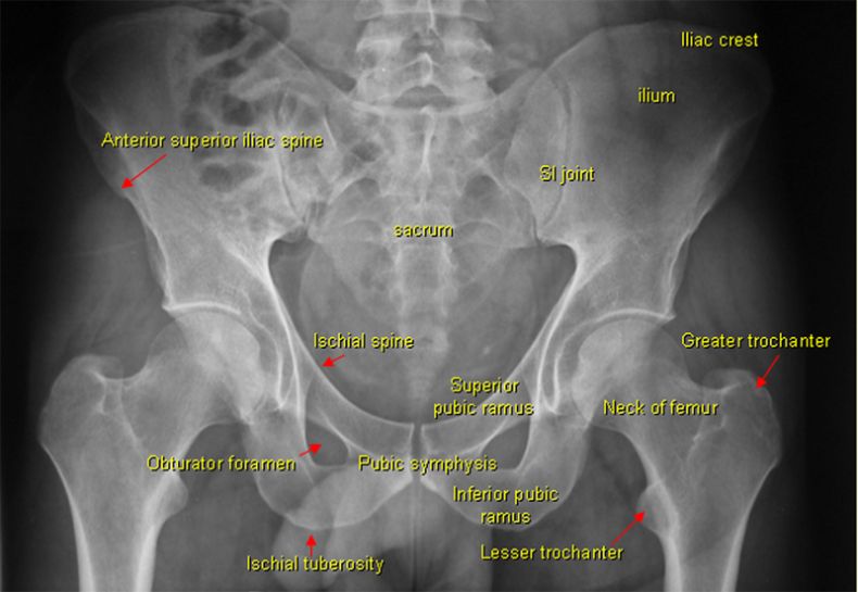
Normal Pelvis Anatomy
Phleboliths
Phleboliths are the most common pelvic calcification. Phleboliths tend to have a characterstic distribution, size and shape. They can sometime cause difficulties in patients who present with renal colic. Finding a renal stone poses a challenge in a sea of phleboliths. Philip Shemilt from Salisbury General Hospital noted the following about phleboliths.
| Phleboliths originate as thrombi in the pelvic veins and are the result of injury to the vein wall. Although in children the pelvic veins and venous plexuses may be protected by valves, these have disappeared in adult life. In 20 dissections in adult cadavers remnants of osteal valves were sometimes seen, but a functioning bicuspid valve was noted only once. These unprotected veins are subjected to large and abrupt rises in pressure during normal acts of coughing and straining. Because of this the veins are distended and damage to the intima with the formation of a protective thrombus can occur. A locally high concentration of phosphatase in the perivesical tissues would explain why these thrombi become calcified in the pelvis and why phleboliths are so rarely found elsewhere. | ||
| <a class="external" href="http://www3.interscience.wiley.com/journal/112191009/abstract?CRETRY=1&SRETRY=0" rel="nofollow" target="_blank">http://www3.interscience.wiley.com/journal/112191009/abstract?CRETRY=1&SRETRY=0 </a> |
</a>
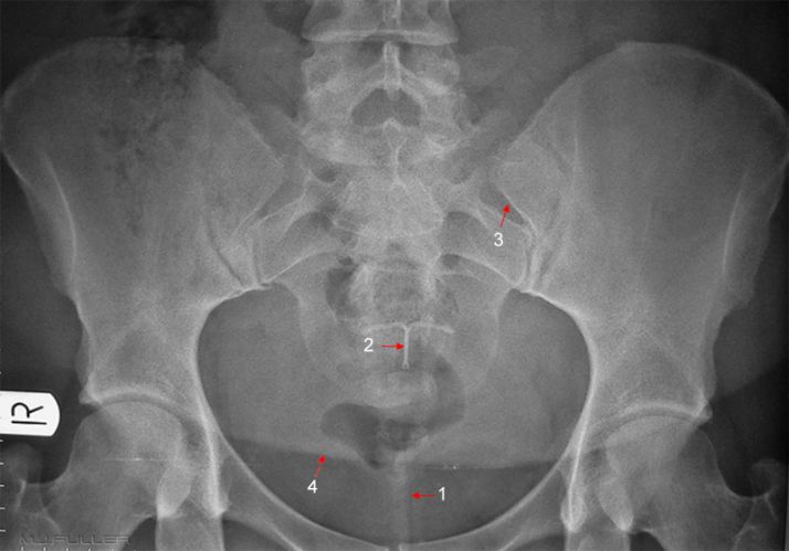
1. Gluteal air (linear dark artifact)
2. Mirena intra-uterine device (IUD)
3. transitional features of transverse processes of L5/S1
4. Abdominal fat "apron"
Case 2
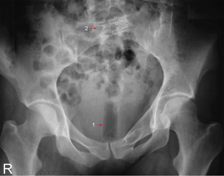
Case 31. Tampon Artifact
2. bra hardware
DiscussionThe tampon artifact is very distinctive in shape and position and rarely causes a problem in terms of misidentification. The bra artifacts are the "eyes" from a "hook and eye" bra fastener. Given that the hook part is not visualised, the bra must be undone (which explains why it appears in the pelvis).
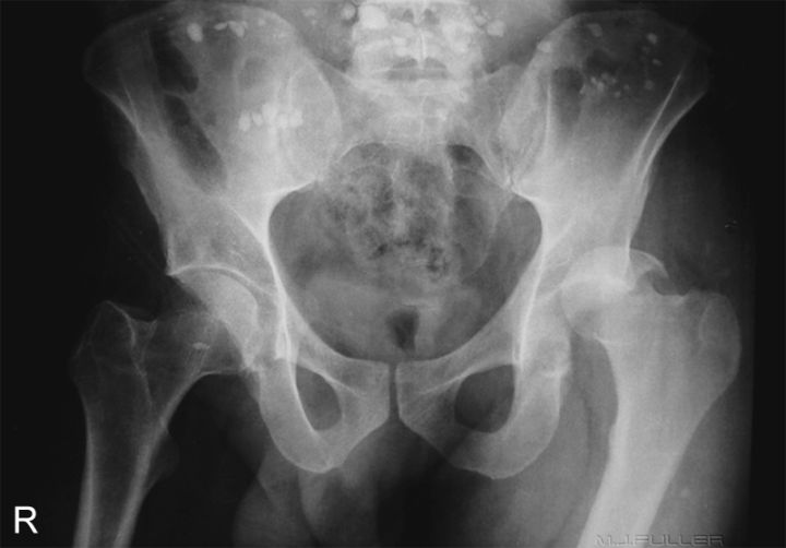
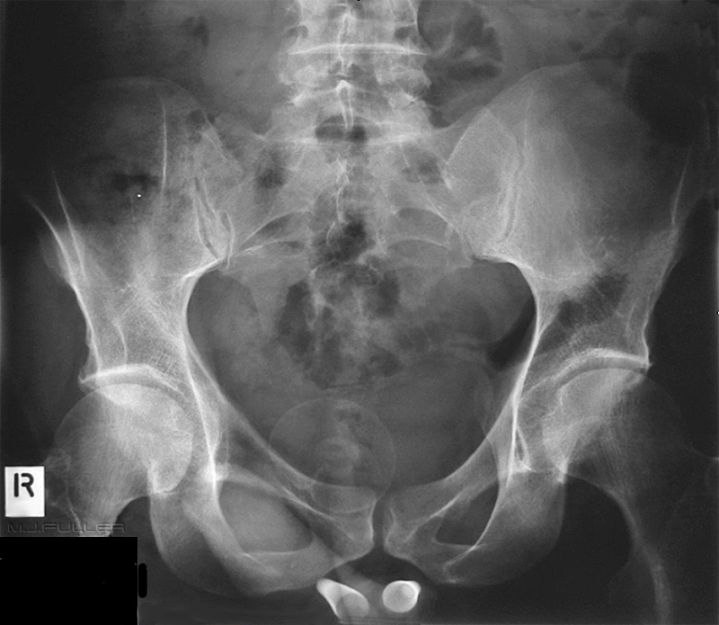
Why do the obturator foramen appear asymmetrical? Is this due to fracture, old healed fracture or is the appearance positional?
This patient presented to the Emergency Department following a motorbike accident (motorbike vs kangaroo). The numerous artifacts dispersed across the upper part of the image are almost certainly small stones from the road that were picked up with the patient by the ambulance officers at the accident scene.
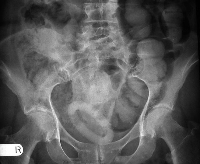
Can you see any bony pathology?
Case 4
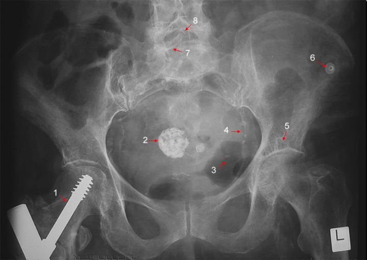
Case 51. hip screw
2. calcified fibroid
3. unidentified curvilinear artifact
4. calcification in iliac artery wall
5 ? small geode
6. ECG dot
7. right lamina- ? partial spinabifida occulta
8. surgical staples
Case 6Penile implants.
For further information
Richard H. Cohan, N. Reed Dunnick, and Culley C. Carson
Radiology of Penile Postheses
AJR:152, May 1989 pp925 - 931
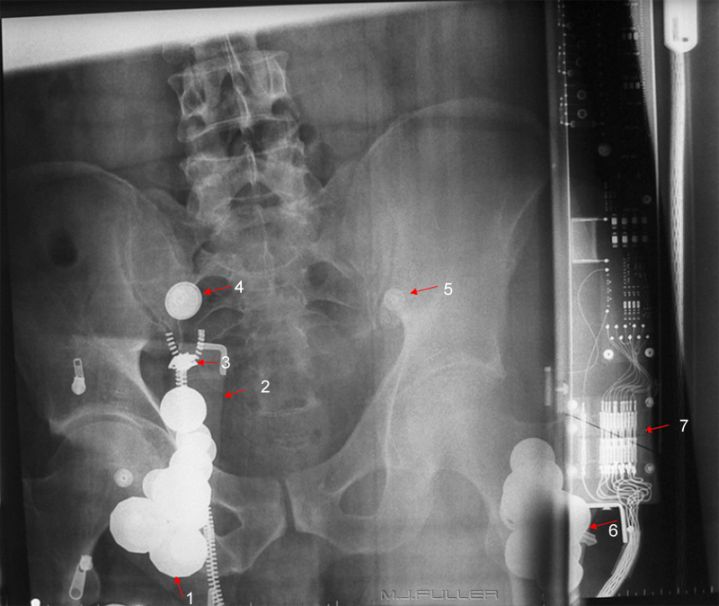
Case 7This is an AP pelvis image on a patient who has presented to the Emergency Department following a motor vehicle accident. The X-ray tube appears to not be centred to the bucky/grid causing grid cut-off artifact. In addition the following artifacts are demonstrated
1 coins
2 wallet
3 zipper
4 jeans stud
5. ?ECG dot ? placement
6. key
7. bucky electronics
The ring opacity is a pessary. Google search "pessary" for more information
Case 8
Case 9
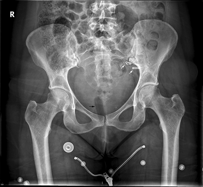
There is a tampon artifact (black arrow)
There are artifacts from metal jean studs and a zipper artifact.
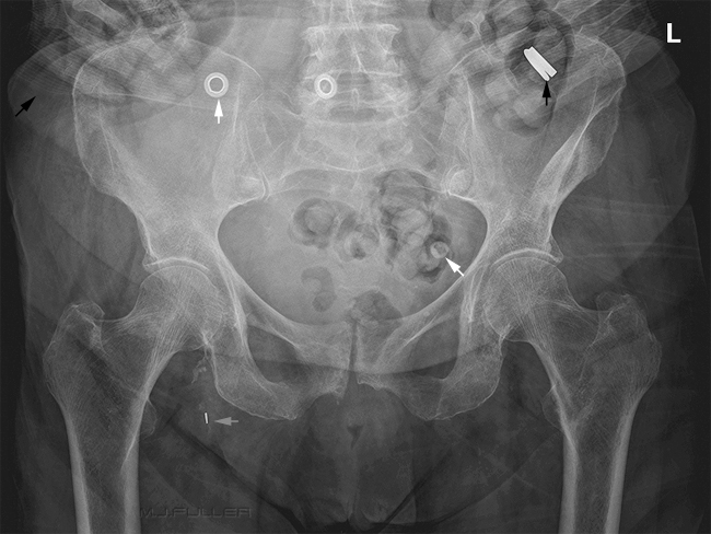
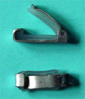
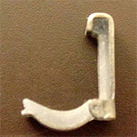
The arrowed metalic artifacts are probably Filshie clips
<a class="external" href="http://www.kugener.com/museum-sub.php?subkategorie=tubar&show=5" rel="nofollow" target="_blank">http://www.kugener.com/museum-sub.php?subkategorie=tubar&show=5</a>
<a class="external" href="http://www.kugener.com/museum-sub.php?subkategorie=tubar&show=5" rel="nofollow" target="_blank">http://www.kugener.com/museum-sub.php?subkategorie=tubar&show=5</a>Filshie Clip Hulka Clip
....back to the Applied Radiography home page here
