Pelvic Calcifications
Introduction
The pelvis is a fertile area for calcifications visualised on plain film. This page provides examples of common pelvic calcifications. There is a related page on normal anatomy and pelvis artifacts.
Case 1
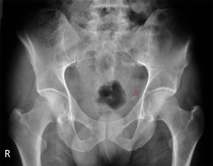
Case 2The larger calcified arrowed structure is almost definitely a phlebolith which is a calcific ring in the wall of a pelvic vein. The smaller calcific structure is also probably a phlebolith but has some of the features of a renal stone in terms of its position and shape. This patient presented after trauma and had no history of renal colic- this mitigates against a renal stone.
Phleboliths are most commonly seen in the lower pelvis and sometime occur in large numbers. They are of no particular pathological significance. Given that they are located in the wall of a vein, they tend to take on characteristics of vein wall- that is, they tend to be:
- round in shape (but not always)
- of a similar size that would correspond to the diameter of pelvic veins
- can look like a ring of bone
- can occur in clusters
- can occur in symmetrical patterns
- tend to occur laterally around the urinary bladder
Phleboliths can be difficult to distinguish from renal stones in the distal ureter. It is useful to consider their size, shape and position. It is also helpful to be aware of the patient's clinical presentation and history. Furthermore, old imaging can be very instructive- a calcified structure that looks like a renal stone might appear in a very short time-frame, whereas a phlebolith will persist over a long period of time.
NB this patient presented after trauma- can you see the 3 fracture(s) of the pelvis. Search this wiki for the case study
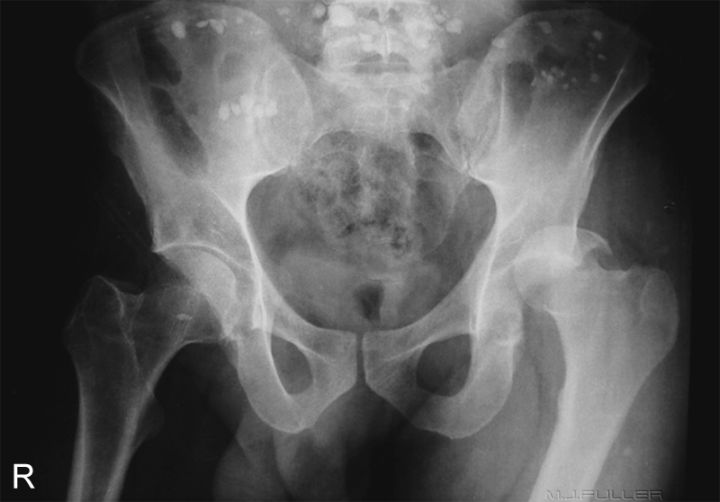
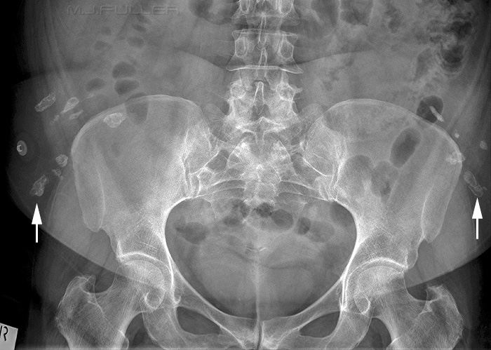
This patient presented to the Emergency Department following a motorbike accident. The numerous artifacts dispersed across the upper part of the image might look like calcific densities but are almost certainly small stones. This patient was found on the roadside following a motorbike accident and the stones were probably picked up with the patient by the ambulance officers at the accident scene.
Can you see any bony pathology?
Case 3
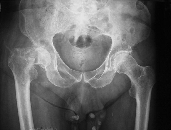
The calcified lesions at the bottom of the image are scrotal calculi which are also known as a fibrinoid loose bodies or scrotal pearl. These structures are contained within the scrotum and may be associated with repetitive trauma such as that which mountain bikers may experience.
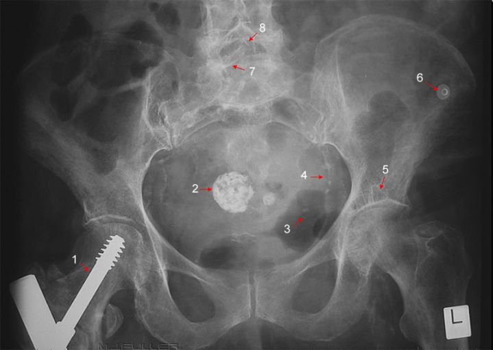
Case 51. hip screw
2. calcified fibroid
3. unidentified curvilinear artifact
4. calcification in iliac artery wall
5 ? small geode
6. ECG dot
7. right lamina- ? partial spina bifida occulta
8. surgical staples
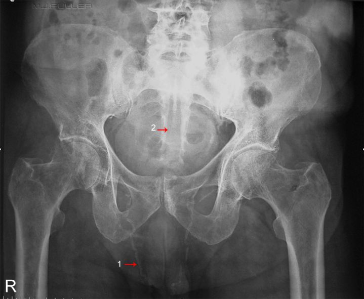
Case 61. Calcified vas deferens (also called the ductus deferens) [thankyou xiongyi]
2. missing posterior sacral elements/ spina bifida occulta
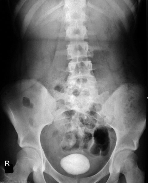
Case 7This patient has a large bladder stone. Bladder stones are uncommon but not rare findings. Note that this stone has a faint longitudinal lucency which is the nidus around which the stone developed. The stone was removed surgically.
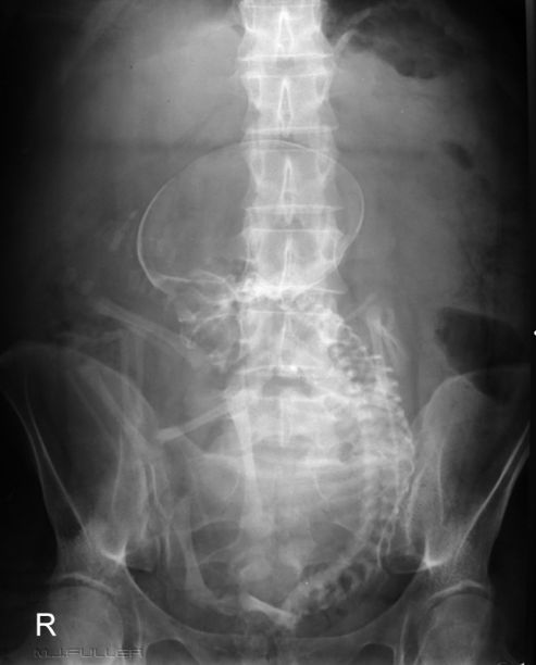
If you haven't noticed the extensive abdominal calcification in this image, it's probably time for a career change! Abdominal images containing a pregnancy are rarely seen today. This abdominal image is circa 1970's. These images were taken to assess foetal position and foetal maturity before the development of ultrasound imaging. Note the breach presentation.
Case8
....back to the applied radiography home page here
