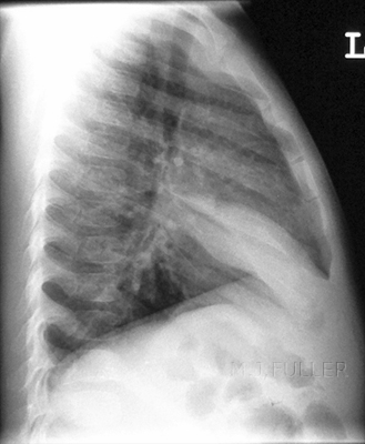
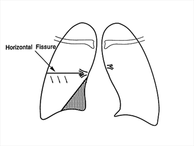
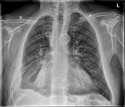
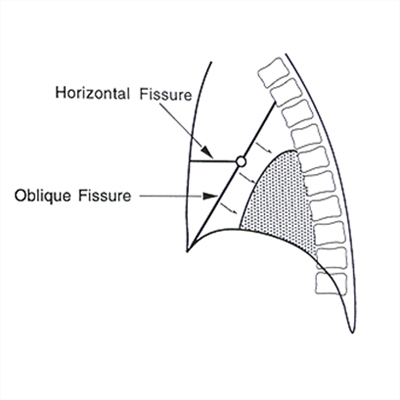
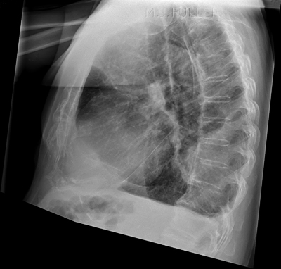
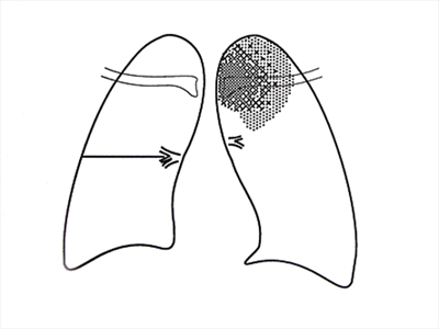
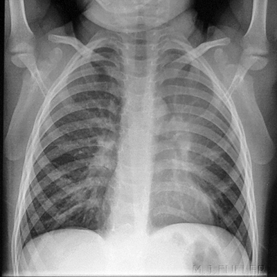
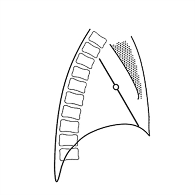
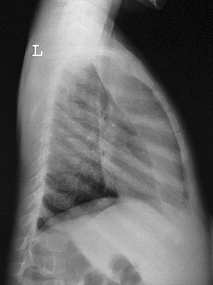
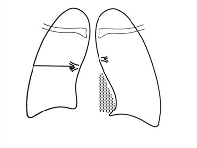
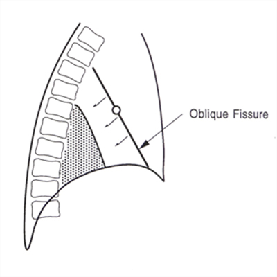
Patterns of Collapse
Definition of TermsFor those with an interest in plain film image interpretation, patterns of collapse and consolidation are a very good place to start learning. Plain film chest interpretation is something of a holy grail. It might appear to be too difficult to contemplate, but as with most seemingly insurmountable tasks, if you take it a step at a time, you will succeed. This page provides an introduction to the topic.
Lung collapse refers to the loss of normal aeration and associated loss of volume(akin to deflating a balloon). The term consolidation when used by a radiologist, refers to the displacement of the air in the alveoli, smaller bronchii, and bronchioles, by exudate or oedematous fluid (Sutton, 1975, p325)
Anatomy
Patterns of collapse and consolidation can definitely not be learned without learning lung anatomy first.
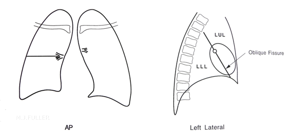
Left Lung
The left lung has •1 fissure•2 lobes
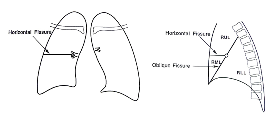
Right Lung
The right lung has •2 fissures•3 lobes
Collapse
Plain Film Appearances of Lung Collapse
Radiological appearances common to all lobes are:
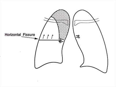
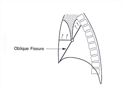
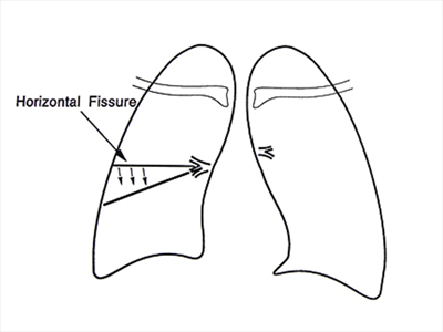
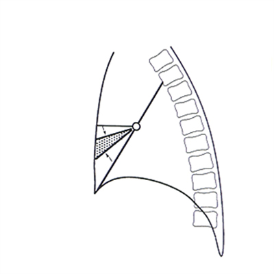
1. Reduced volume of the lobe2. Opacity of the lobe3. Displacement of fissures towards the opaque lobe4. Vascular shadows in the remainder of the lung are more widely spread than normal5. Hilar/mediastinal/diaphragmatic/tracheal displacement
Dixon and Dugdale, 1988,p25Notes1. Collapse and consolation can occur independently or together2. Collapse can be partial or complete3. It is often not clear to what extent the appearance is due to collapse or consolidation or both. The degrees of each are often unclear.4. If a lobe is only partially collapsed and there is no accompanying consolidation, there may be no increase in opacity5. In cases of pure collapse, only when the collapse is virtually complete will there be a significant increase in density of the affected lung
Hilar DisplacementHilar displacement is an important indirect sign of lobar collapse. In most patients the left hilum is slightly higher than the right. Elevation of the hilum can be seen in cases of upper lobe collapse and depression of the hilum may be seen in lower lobe collapse. Middle lobe and lingular collapse do not affect the hilar positions.
Right Upper Lobe (RUL) Collapse
Right Middle Lobe (RML) Collapse
Right Lower Lobe (RLL) Collapse
Left Upper Lobe (LUL) Collapse
Left Lower Lobe (LLL) Collapse
...back to the Applied Radiography home page
