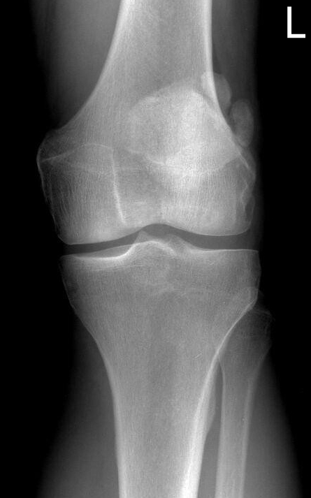
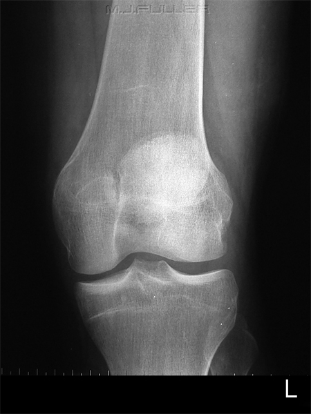
IntroductionPatellar fracture and dislocations are commonly seen by radiographers in the Emergency Department. They are most likely to be missed because of radiography failure (usually failure to include skyline projection in the series) and/or interpretation failure (fracture interpreted as normal variant and vice versa). This page considers all aspects of plain film radiography of patellar fractures and dislocations.
Other Wikiradiography Pages of InterestThe Skyline Patella Projection
Soft Tissue Signs- Knee Trauma
Soft Tissue SignsSubcutaneous Soft Tissues
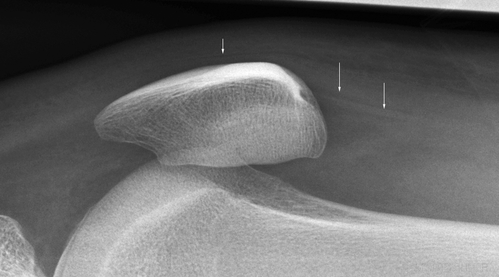 |
| It can be difficult to distinguish between a patellar fracture and a multipartite patella. When considering the possibility of patellar fracture in htis patient, note that there is clear delineation of the fat and fluid density subcutaneous soft tissue planes. |
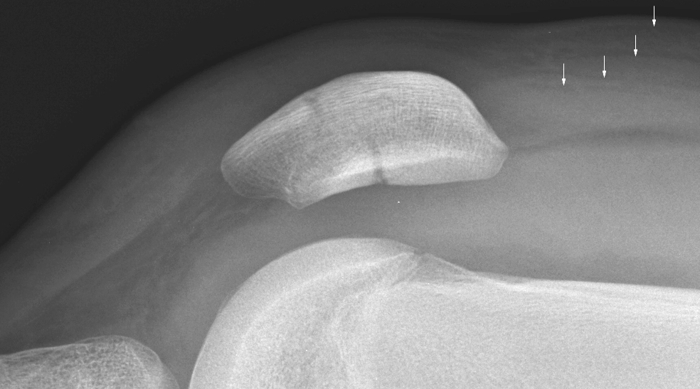 |
| This patient demonstrates clear delineation of the subcutaneous and deeper soft tissues superior to the patella. The tissues overlying the mid-patella demonstrate loss of definition (mixed fat/fluid density) and there is soft tissue swelling overlying the lower pole of the patella. In conjunction with a large knee effusion and a bony patellar defect, this would suggest that the patient has a patellar fracture. |
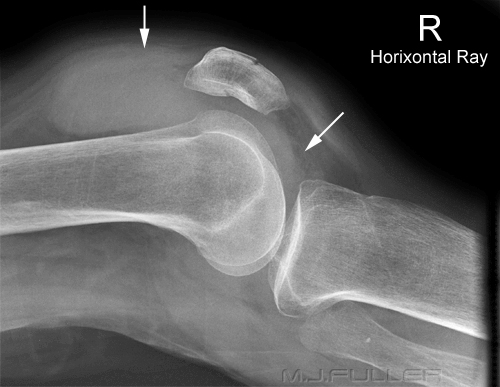 | A fractured patella is usually a ostochondral fracture which will, by definition, communicate with the knee joint. It is common for these fractures to be associated with a knee joint effusion which will be best demonstrated on the lateral knee projection image. A knee joint lipohaemarthrosis may also be demonstrated.
The patient has a fractured patella and an associated large knee joint effusion (arrowed) |
|
|
|
|
|
|
Is the Skyline Projection Necessary?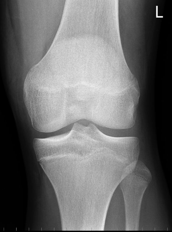 | This 17 year old male presented to the Emergency Department following a fall from his bike. He was examined and the requested radiography included his left knee.
Is there any evidence of fracture or dislocation? |
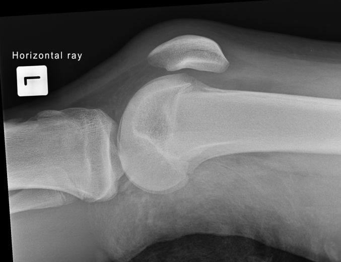 | The lateral knee projection image demonstrates no fracture or dislocation.
Is further imaging required? |
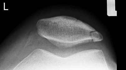 | The skyline projection image demonstrates a possible fracture of the lateral aspect of the patella. Is this a fracture or a secondary ossification centre? If you scroll up the page and review the AP image you will notice that there is a faint impression of a lucency over the supra-lateral aspect of the patella- this is the most common position to find a secondary ossification centre. If this was a fracture, would it have been missed without the skyline projection included in the routine radiographic series? |
Acute Fracture, Old Fracture or Normal Variant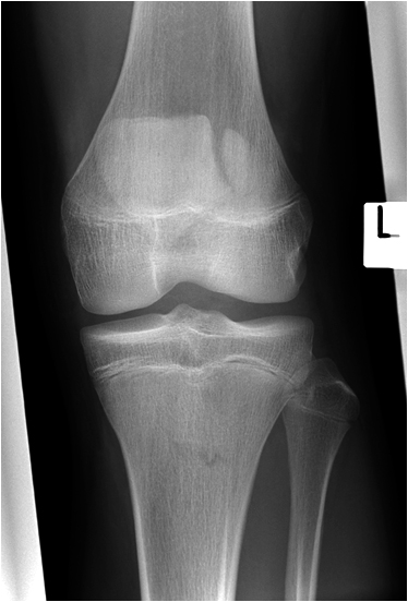 |
|
This patella has a number of features suggesting that it is bipartite rather than fractured
- The position of the 'fragment' is supero-lateral
- The fragment margins appear smooth
- The fragment will not fit back (like a broken biscuit) to make a normal smooth contoured patella
|
|
|
|
|
|
Case StudiesCase 1
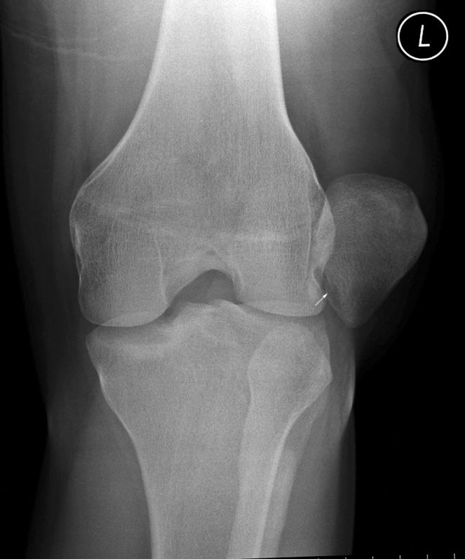 | This 23 year old male presented to the Emergency Department with a dislocated left patella. This is somewhat unusual in that patients who experience a dislocated patella will often relocate the patella themselves prior to presentation to the Emergency Department.
The AP knee projection image demonstrates the patella to be dislocated laterally. There appears to be a lucent defect in the inferomedial aspect of the patella. This may represent an avulsion fracture. |
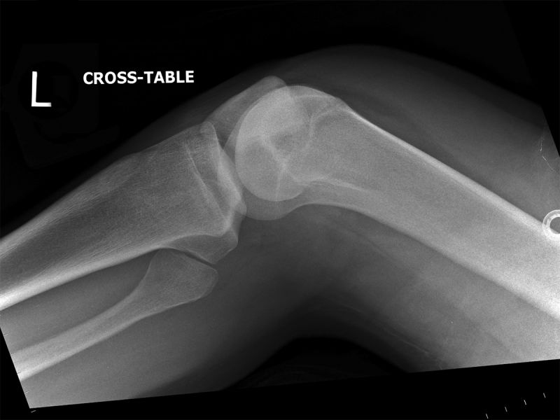 | The cross-table lateral knee image demonstrates the dislocated patella. |
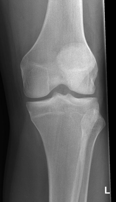 | The patella was relocated in the ED. The post-relocation AP knee image demonstrates the relocated patella. The patella is not centrally located due to external rotation positioning error. |
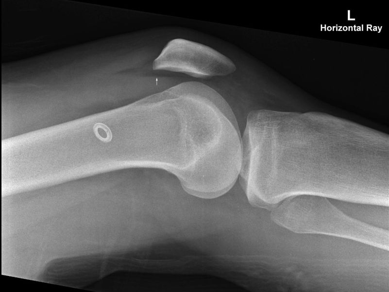 | The post-relocation lateral knee projection demonstrates the relocated patella and a lipohaemarthrosis (arrowed). The lipohaemarthrosis supports the possibility that the patient suffered an avulsion fracture of the patella with the donor site demonstrated on the pre-reduction AP knee image. |
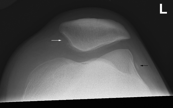 | The post-reduction skyline patellar image demonstrates the patella to be relocated. The possible site of the avulsion fracture is shown (white arrow). A bony fragment (black arrow) demonstrated on the lateral aspect of the lateral femoral condyle is of unknown significance. |
Case 2
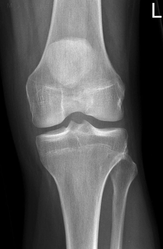 | This 34 year old male presented to the Emergency Department following a fall from his pushbike. On examination, he was found to have a painful and swollen left knee. He was referred for knee radiography.
The AP projection image demonstrates patella alta. Patella alta refers to a condition in which the patella is located in an abnormally proximal position. |
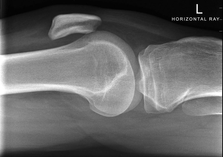 | The cross-table lateral knee image also demonstrates patella alta.
Skyline knee radiography will be difficult given the high-riding psoition of the patella. It may be necessary to flex the patient's knee more than would be normally required in order to project the tibial tuberosity clear of the patello-femoral joint. |
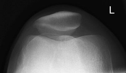 | The skyline projection demonstrates tibial tuberosity overlying the patello-femoral joint space. |
Case 3
| | This patient presented to the Emergency Department after a fall onto her left knee. The patient was referred for a knee X-ray examination. The patient's patella was tender but there was very little soft tissue swelling.
There appears to be a vertically orientated lucency through the medial aspect of the patella. This could represent a new fracture, an old fracture, or a bipartite patella.
This is an uncommon site for a bipartite patella (more commonly sited supero-lateral). |
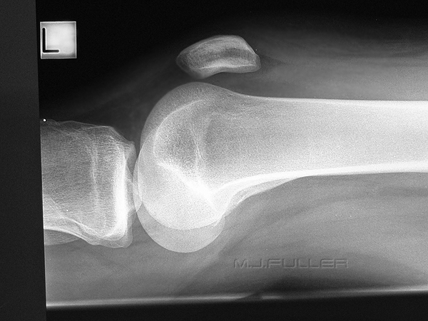 |
There is possibly a very small joint effusion (see Hoffa's fat pad and the suprapatellar pouch)
There is no obvious soft tissue swelling overlying the patella but the image is a little overexposed for the soft tissues (pre-digital image) |
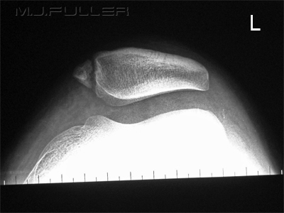 |
There is evidence of a corticated margin to the "fracture" suggesting that it is not an acute injury. |
|
|
....back to the Applied Radiography home page here


