Paediatric Wrist Trauma
Jump to navigation
Jump to search
The physis or Growth Plate
Imaging
Discussion
Radiographic Technique
Referral
Image Evaluation
Discussion
Presentation
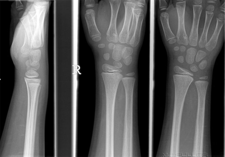
This nine year old boy presented to the Emergency Department (ED) following a fall onto an outstretched hand. Following a history/clinical examination by the doctor in the ED, he was referred for a wrist X-ray examination.
Clinical Information Provided
“FOOSH. Swelling anteriorly over radius. No obvious deformity”
The physis or Growth Plate
The distal radial physis fuses at approximately 20 years. During, as well as after the fusion, the "scar"of the physis may simulate a fracture. (Harris, H.H., Harris, W.H., and Novelline, R.A. The Radiology of Emergency Medicinem 3rd Ed., 1993, p379)
Imaging
Discussion
It is a good practice for a trauma radiographer to confirm the clinical information that is provided by the referring doctor directly with the patient (where appropriate). This may help clarify relative terms such as pain, swelling and deformity (…how much pain? how extensive? is it new? is it related to the injury?). Significant additional relevant information may also be revealed. This approach will assist the Radiographer to approach the examination with the confidence that follows from fully understanding what you are doing and why you are doing it.
FOOSH is an acronym for Fall Onto OutStretched Hand and is a common mechanism of injury, particularly in children and the elderly. The most common bony wrist injuries in children are the greenstick and buckle/torus fractures of the radius.
Radiographic Technique
The PA, lateral and oblique radiographs will demonstrate most bony injuries of the wrist. These images should be examined carefully with a view to performing supplementary views if required.
Referral
You can't expect to achieve good diagnostic standards with the wrong images. The following is an eloquent quote from John Harris on the subject.
| "Anatomically and radiographically, the forearm is not the wrist and the wrist is not the hand. Therefore, it is important for attending physicians to indicate precisely the anatomical part to be examined radiographically. Specifically, the attending physician must define, on the basis of history and physical examination, whether the forearm, wrist, or hand, is to be examined, since the routine views of one of these anatomic areas is of limited practical value with respect to the others. In spite of this admonition, it is important to remember that injuries of the distal forearm may cause, or be associated with, carpal injuries. Finally, contrary to an often-repeated dictum, it is not necessary to radiographically examine both the wrist and the elbow in the instance of a suspected radial or ulnar shaft fracture in a patient who is alert, responsive, and communicative and on whom a proper physical examination can be performed. In patients who do not fulfil these criteria, it is prudent to examine both the wrist and the elbow. In a similar vein, it is not necessary to routinely obtain radiographs of the normal contralateral wrist in children simply 'for comparison'. Physicians interpreting skeletal radiographs of children and adolescents should be sufficiently familiar with the appearance of normal growing bones to be able to determine whether the wrist is normal or not. Only if the examination of the injured pediatric or adolescent wrist is ambiguous should the opposite (normal) side be examined for comparison." | ||
| John Harris, William Harris, Robert Novelline. The Radiology of Emergency Medicine, Third edition. Williams and Wilkins, 1993 | ||
Image Evaluation
There are four areas of interest that should be initially considered in paediatric patients who may have a distal forearm/wrist fracture.
- Are the bony cortices intact? The cortices of the metaphysis and diaphysis should appear as smooth even lines.
- Is the trabecular pattern normal? In particular, is there evidence of a transverse sclerotic band in the distal radius or any abnormal bony lucency?
- Is the volar tilt within normal limits? i.e. does the distal radius look like it is bent backwards (dorsally angulated)?
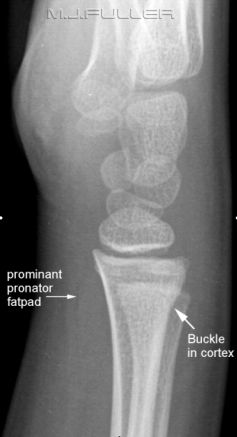
- Does the pronator fat pad suggest a recent significant wrist injury?
.- The dorsal cortex of the distal radius can be seen to deviate from a smooth curve suggesting a buckle fracture. Note that this is very subtle finding but was noted (and 'red dotted') by the radiographer at the time
- A subtle lucent bony defect can be seen in the distal radius closely related to the buckled radial cortex
- Subtle volar tilt or palmar inclination cannot easily be measured in this age group. There does not appear to be significant dorsal angulation of the radius
-The pronator fat pad is raised suggesting a significant wrist injury (bulging of the fascial covering of the pronator quadratus muscle caused by haematoma)
-A comparison view of the patient’s contralateral wrist is not required to arrive at a diagnosis
.
Diagnosis
Buckle fracture of the distal radius with no significant dorsal angulation.
Discussion
....back to the applied radiography home page hereIdentifying these types of subtle buckle fractures is within the diagnostic capabilities of a radiographer. In my institution they are often picked up by the radiographer and the findings relayed to the referring doctor via the electronic PACS based Red Dot System. Such cases are reviewed by the radiographers every fortnight at the radiographer's Case Review Meetings. This allows the experienced radiographers to share their knowledge and for all radiographers to discuss the radiographic techniques employed in displaying particular pathologies.
You will hear radiographers dismiss this type as finding as 'unimportant' because"...it does not affect the patient's management"
What they are suggesting is that the patient would not have had operative treatment whether the fracture was detected or not. This reflects a limited understanding of the patient experience and how a patient is managed and treated. It may also reflect an outmoded photographic approach to radiography rather than a more appropriate clinical approach.
If the fracture is detected, the attending doctor is able to explain to the patient/parent that the patient's symptoms have been accounted for. This is comforting for the patient/parent. The doctor can explain to the patient what to expect in the following weeks in terms of associated limitations of movement and pain. He/she is also able to prescribe an appropriate level and type of pain relief and apply appropriate immobilisation (plaster/backslab/fibreglass rather than sling or nothing). A full plaster or backslab will reduce pain and decrease the likelihood of re-injury.
It is well worth developing a holistic view of your role as a radiographer- one that reflects your role as a part of a health care team rather than being limited by a photographic based picture-centric view. You will find your role more satisfying if you extend you thinking beyond that of a "medical photographer".

