OPG
Jump to navigation
Jump to search
An OPG which stands for ORTHOPANTOGRAM, gives a panoramic view of the mandible and teeth.
It is performed using a technique called "tomography". The X-ray tube moves around the head, the x-ray film moves in the opposite direction behind your head. This generates an image slice where the mandible and teeth are in focus, and the other structures are blurred.
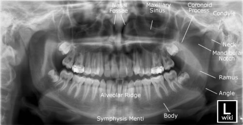
Anatomy
The anatomy consists of the body, ramus and angle of mandible, coronoid process, mandibular notch, condyle of mandible, alveolar ridge, symphysis menti, maxillary sinuses, nasal fossae and 16 upper and 16 lower teeth, as shown in the image below.
It is performed using a technique called "tomography". The X-ray tube moves around the head, the x-ray film moves in the opposite direction behind your head. This generates an image slice where the mandible and teeth are in focus, and the other structures are blurred.
Anatomy
The anatomy consists of the body, ramus and angle of mandible, coronoid process, mandibular notch, condyle of mandible, alveolar ridge, symphysis menti, maxillary sinuses, nasal fossae and 16 upper and 16 lower teeth, as shown in the image below.

Palmer Notation
The mouth is divided into four sections called quadrants (upper and lower) right quadrant and (upper and lower) left quadrant. The numbers 1 through 8 are used to label each of the teeth in all four quadrants. The numbering runs from the center of the mouth to the back as shown in the image below.
The mouth is divided into four sections called quadrants (upper and lower) right quadrant and (upper and lower) left quadrant. The numbers 1 through 8 are used to label each of the teeth in all four quadrants. The numbering runs from the center of the mouth to the back as shown in the image below.
<img align="bottom" alt="OPG - wikiRadiography" height="253" src="http://image.wikifoundry.com/wiki/wikiradiography/image/119+U2LswMfYYU+CvhF9IGg==71441/GW500H253" title="OPG - wikiRadiography" width="500"/>
In Adults there are 32 teeth. Teeth numbered 1&2 in each quadrant are called incisor. The third tooth is called canine. Numbers 4&5 are called premolar. Numbers 6,7&8 are called molar. The 8th tooth (3rd molar) are commonly called wisdom teeth.
Below is a Pediatric OPG showing the adult teeth above the gum line.
Reasons for OPG requests
Below is a Pediatric OPG showing the adult teeth above the gum line.
<img align="bottom" alt="OPG - wikiRadiography" height="257" src="http://image.wikifoundry.com/wiki/wikiradiography/image/1Vg23q$7KISGCr$mOVgLVSg==36362/GW500H257" title="OPG - wikiRadiography" width="500"/>
Reasons for OPG requests
Dental Disease
- Caries - appear as different shaped areas of radiolucency in the crowns or necks of teeth.
- Peridontioiditis - when inflammation extends into the underlying alveolar bone and there is a loss of attachment.
- Peridontal Abscess - Radiolucent area surrounding the roots of the teeth.
Extraction of teeth (eg. wisdom teeth)
- OPG shows angulation, shape of roots, size and shape of crown, effect on other teeth.
Teeth Abnormalities
- Eg. Developmental, to show size, number, shape and position.
Trauma to teeth and facial skeleton
- Mandible fractures are often bilateral.
- Panoramic view of mandible to view the fracture.
- Determine site and direction of fracture lines.
- Relationship of teeth to fracture lines.
- Alignment of bone fragments after healing.
- Evidence of infection or other complications post intervention.
- Follow up to assess healing.
Transplant workup
- To look for evidence of any underlying dental disease (eg. abscess)
- Patients on steriods after a transplant are immunosuppressed and the mouth is a common site of infection.