Notes on Paediatric Chest Radiography

From wikiRadiography
Jump to navigation Jump to search
Introduction

Paediatric chest radiography tends to be done very well in dedicated paediatric institutions and less well everywhere else. It is worth arranging to spend some time in a paediatric imaging department to develop paediatric radiography skills. This page covers all aspects of paediatric chest radiography technique.

Relevant Wikiradiography pages

Notes on Paediatric Chest Radiography

  • Children over the age of 5 years are usually able to be positioned in a PA erect position for chest radiography. (some 4 year olds will be sufficiently compliant for PA/AP erect chest radiography)
  • There is very little difference between PA and AP chest radiography in children (Caffey 1978, and Hochschild and Cremin, 1975)
  • There is very little difference between left and right lateral chest radiography in children


  • AP Chest radiography has several advantages over PA chest radiography in children
    • The radiographer can watch the child's breathing in order to expose on full inspiration
    • It is easier to hold a child straight and still
    • The child can watch the tube being centred and the beam collimated without turning around
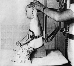
  • The child's arms should be held against the side of the child's head with elbows flexed and pointing forwards.
  • an 18 x 24cm is suitable for children up to about 2 years
  • an 24 x 30cm cassette is suitable for children up to about 7 or 8 years
  • movement of the abdomen rather than the chest is watched to ensure exposure on full inspiration
  • Exposure time should be less than 0.02 seconds
  • A paediatric lead rubber apron should be employed for gonad protection for the child
  • The adult holding the child should be protected by at least 0.5mm lead equivalent

(Catherine Gyll, Noel Blake, and A Thornton. Paediatric Diagnostic Imaging, 1985, p4,John Wiley and Sons, New York)

  • It is undesirable to position the child/baby with the legs horizontal for chest radiography- some kind of seat is preferred such that the child's hips are higher than his/her knees
  • When examining children over the age of 3 it is useful to have the child practice taking a deep breath before the exposure.

Preventing the Lordotic Projection
paediatric chest radiography
adapted from
Radiography, Preventing Lordotic Projection of the Chest, Catherine Gyll, December 1983, V49, No 588, pp291-293
Catherine Gyll considered the unintentional lordotic positioning error to be of such importance that she wrote an article on the subject. (Radiography, Preventing Lordotic Projection of the Chest, Catherine Gyll, December 1983, V49, No 588, pp291-293)

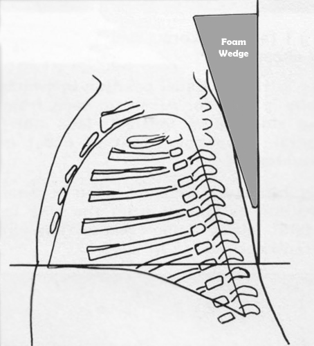
Up to 1/3 of the baby's lung field can be hidden below the diaphragm on AP/PA chest radiography if care is not taken to avoid a lordotic projection. A 15 degree wedge sponge placed behind the baby's chest can assist considerably in avoiding an unintentional lordotic projection.

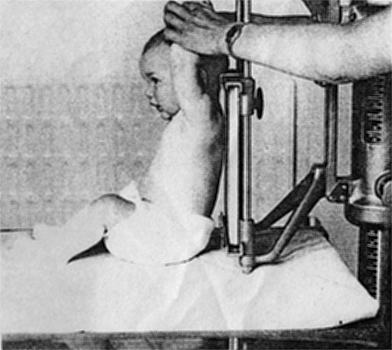
paediatric chest radiographyA baby child will tend to end up in this position when seated with legs horizontal. This position will result in a lordotic projection.
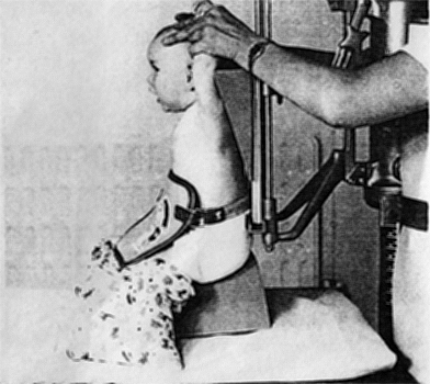
paediatric chest radiographyIf the baby is seated on a small sat and a 15 degree sponge placed behind the child's back, a more satisfactory position is achieved.


Case 1
Notes on Paediatric Chest Radiography - wikiRadiographyThe child is positioned for AP chest radiography in the erect sitting position. The adult who is holding the child's arms is holding them out to the sides (abducted) rather than as shown below. This position allows the child's head to fall forwards and rotate.

paediatric chest radiography
Notes on Paediatric Chest Radiography - wikiRadiographyThe child's arms are correctly positioned for the lateral projection.
Notes on Paediatric Chest Radiography - wikiRadiographyIt is not sufficient to simply hold the child's arms in a raised position. The adult holding the child must use their fingers to position the child's head. This child was slumped with head turned to the right. The patient does not have a scoliotic spine- the appearance is positional


Case 2

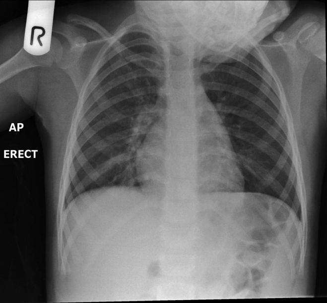
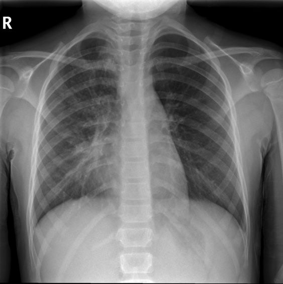
This 6 year old girl presented to the Emergency Department with a temperature of 39 degrees. She was assessed and referred for chest radiography

Notes on Paediatric Chest Radiography - wikiRadiographyNotes on Paediatric Chest Radiography - wikiRadiography
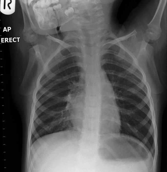
At 6 years of age the radiographer correctly considered that she would be able to stand against the erect bucky for her chest radiography. At this age AP erect radiography has advantages over PA erect radiography. The radiographer noted what appeared to be abnormal right hilar opacity and abnormal opacity if the RUL. The appearance was not easily accounted for given that the most likely cause of the patient high temperature was infection.

The radiographer considered that the unusual appearance of the hilum might reflect malpositioning (unintended lordotic positioning) rather than hilar pathology. The AP erect chest was repeated with the aim of correcting the lordotic malposition
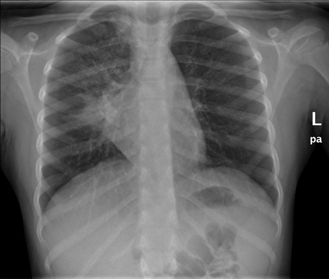
The repeated AP erect position showed considerable improvement- this image is easier to interpret- the patient has a silhouette sign involving the RML and right heart border and abnormal opacity in the RUL (compare with LUL)
Notes on Paediatric Chest Radiography - wikiRadiographyNotes on Paediatric Chest Radiography - wikiRadiography
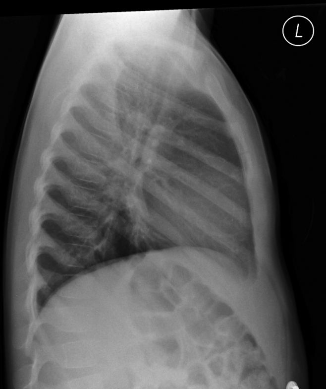
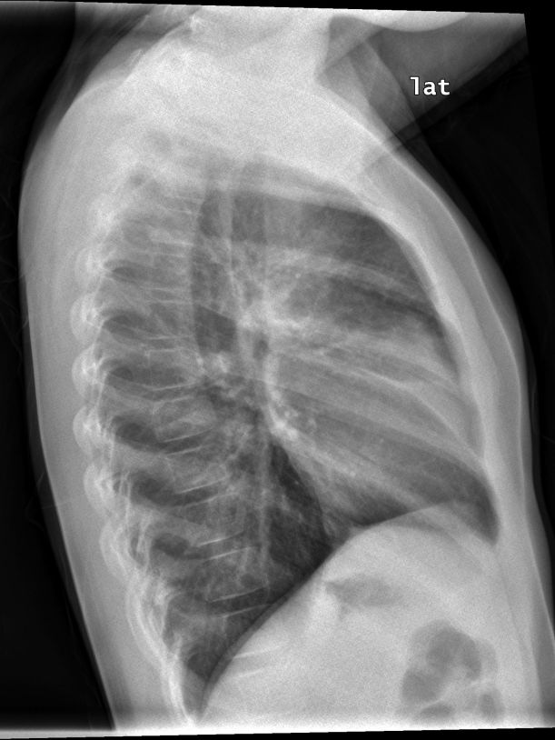
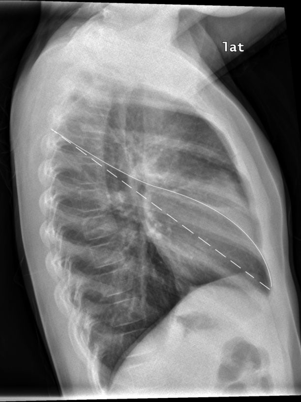
The lateral projection image demonstrated evidence of collapse/consolidation of the RML and RULThe solid line marks the position of the right oblique fissure and the dotted line marks the approximate normal position of the oblique fissure

Comment

This case illustrates the vital link between a radiographer's image interpretation skills and their radiographic skills- proficiency in image interpretation will assist in deciding when to repeat and when not to repeat. The original AP erect chest image demonstrates an abnormal pattern that you would not expect in a child who presents with a high temperature. Common pathologies and patterns of pathology occur commonly and they are looked for first. It was the combination of pattern recognition skills and image evaluation skills that led the radiographer to the conclusion that the patient's pathology had not been demonstrated correctly. The repeat AP erect chest allows for the easy recognition of RML and RUL disease- it is a pattern that is easily recognisable in a correctly positioned AP/PA chest.


... back to the Wikiradiography home page
... back to the Applied Radiography home page