Neonatal Lines, Tubes and Catheters
Introduction
Abbreviations and TerminologyWhen you are summoned to the neonatal unit (commonly known as NNU, SBCU or NICU) for a 'check X-ray', it is often to establish the position of a tube, line or catheter. A knowledge of these lines is useful in producing an image that is appropriately positioned and coned.
Everything in the neonatal unit (NNU) can be abbreviated into a three letter acronym (TLA).
UAC: Umbilical artery catheter
UVC: Umbilical vein catheter
ETT: Endotracheal tube
NGT: Nasogastric tube
TPT: Transpyloric tube
VPS: Ventriculo-peritoneal shunt
CVC: Central Venous Catheter
TPN: Total parenteral nutrition
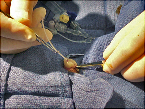
Umbilical Catheter Insertion
Umbilical Vein Catheter Insertion
- Identify and dilate the umbilical vein
- Place line into the lumen and advance to your premeasured goal
- Check to see if the line draws back and flushes
- If feeling resistance or “bouncing” – likely coiled in the liver
Umbilical Artery Catheter Insertion
- Dilate one of the arteries, go for the least tortuous, cut down stump if both are very tortuous
- Insert catheter to calculated length
- If resistance early, 1 to 3 cm, consider side approach or trying other artery
- If resistance further along or no blood return, likely in the leg, try to pull back and reposition or try the other artery
- May also try giving fentanyl for vasospasm
<a class="external" href="http://www.youthclinic.com/Pediatric+lectures/Line+Placement.ppt" rel="nofollow" target="_blank">www.youthclinic.com/Pediatric%20lectures/Line%20Placement.ppt</a>
<embed height="350" src="http://widget.wetpaintserv.us/wiki/wikiradiography/page/Neonatal+Abdominal+Radiography/widget/unknown/086e2cee1e33b89c5ee30df2a69002a3f04f95f7" type="application/x-shockwave-flash" width="425" wmode="transparent"/> <embed height="350" src="http://widget.wetpaintserv.us/wiki/wikiradiography/page/Neonatal+Abdominal+Radiography/widget/unknown/ad00a81210de0ef9818732c447307888e9eb2923" type="application/x-shockwave-flash" width="425" wmode="transparent"/> This video demonstrates the insertion technique for a umbilical catheter in a mannequin This is an instructional video on how to insert an umbilical catheter. The sound quality is awful and is not in English.
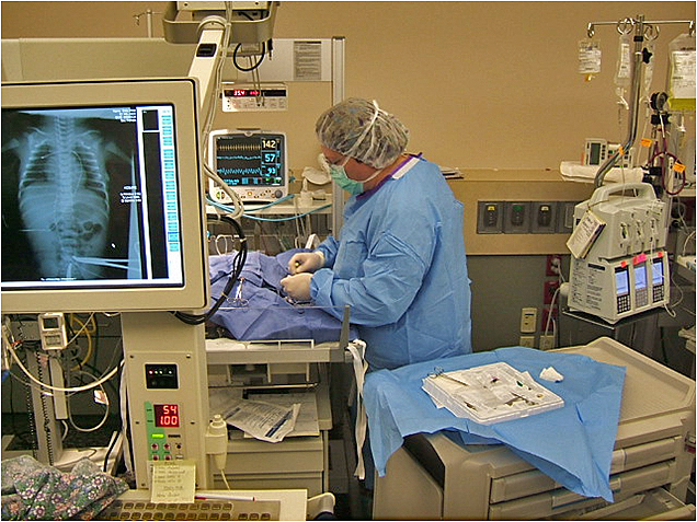
The Case for Checking Lines with DR in NICU
Neonatal Vascular Anatomy
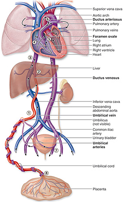
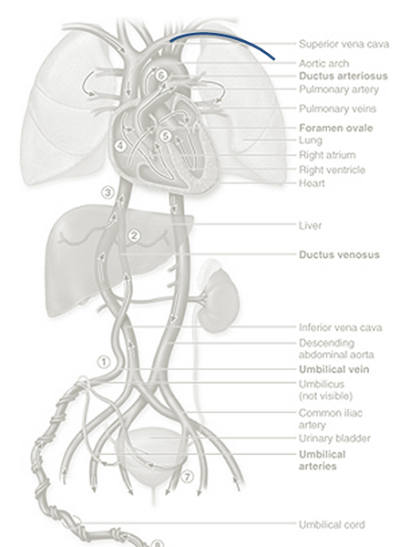
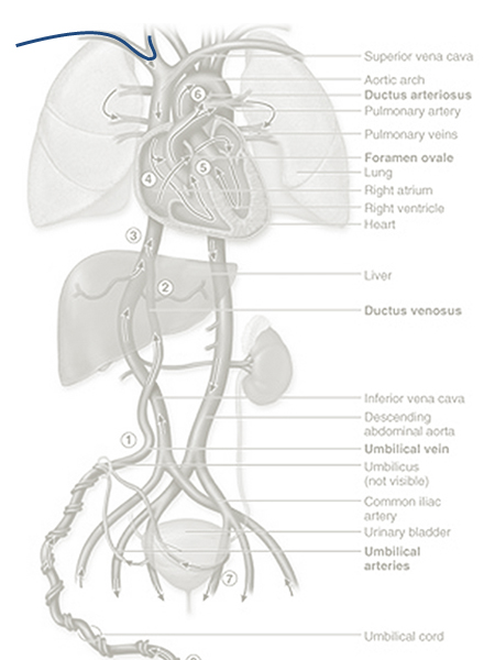
The positioning of umbilical catheters in particular is easier to digest if you understand the foetal vascular anatomy
Umbilical Arterial Catheter (UAC)
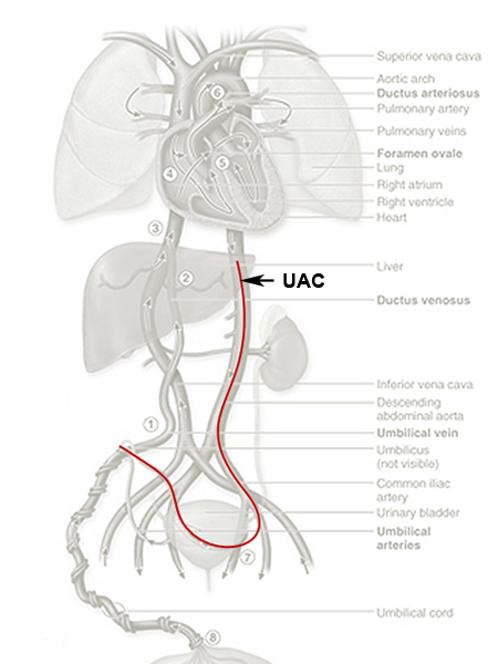
- Placed into the umbilical artery, through to the descending aorta
- Allows continuous blood pressure monitoring
- Allows for frequent blood gas, lab sampling
<a class="external" href="http://www.youthclinic.com/Pediatric+lectures/Line+Placement.ppt" rel="nofollow" target="_blank">www.youthclinic.com/Pediatric%20lectures/Line%20Placement.ppt</a>
Umbilical Vein Catheter (UVC)
- The umbilical arteries are the direct continuation of the internal iliac arteries.
- A catheter passed into an umbilical artery will usually (but not always) enter the aorta via the internal iliac artery.
- Occasionally it will pass into the femoral artery via the external iliac artery or into the gluteal arteries.
- The femoral artery or gluteal artery are unsuitable sites for sampling, infusion, or blood pressure monitoring
<img align="bottom" alt="Umbilical catheter position" height="600" src="http://image.wikifoundry.com/image/1/GWJuIjLId0OcSBW_Q3X4rg131678/GW450H600" title="Umbilical catheter position" width="450"/>
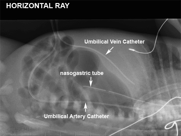
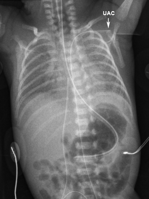
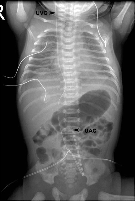
adapted from source: unknownThe umbilical artery catheter (UAC) characteristically deviates inferiorly before tracking up the aorta.
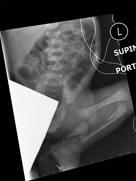
See the lateral decubitus abdominal image below for further appreciation of the course of the umbilical artery.
UAC in High Position
<img align="bottom" alt="Umbilical catheter position" height="600" src="http://image.wikifoundry.com/image/1/GWJuIjLId0OcSBW_Q3X4rg131678/GW450H600" title="Umbilical catheter position" width="450"/>
adapted from <a class="external" href="http://www.youthclinic.com/Pediatric+lectures/Line+Placement.ppt" rel="nofollow" target="_blank">www.youthclinic.com/Pediatric%20lectures/Line%20Placement.ppt</a>
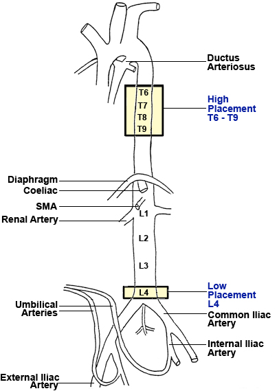
adapted from <a class="external" href="http://www.youthclinic.com/Pediatric+lectures/Line+Placement.ppt" rel="nofollow" target="_blank">www.youthclinic.com/Pediatric%20lectures/Line%20Placement.ppt</a>There are two potential tip positions for the UAC. These are described as "high" or "low".
- The high position is at the level of thoracic vertebral bodies T6-T9. This position is above the coeliac axis (T12), the superior mesenteric artery (T12-L1), and the renal arteries (L1). This position is essentially "above the diaphragm".
- The low position is at the level of lumbar vertebral bodies L3-L4. This position is below the structures as above, and is above the aortic bifurcation (L4-L5). The inferior mesenteric artery arises from L3-L4. This position is essentially "above the aortic bifurcation". <a class="external" href="http://www.adhb.govt.nz/newborn/Guidelines/VascularCatheters/UmbilicalCatheters.htm#UAC" rel="nofollow" target="_blank">source</a>
<a class="external" href="http://www.adhb.govt.nz/newborn/TeachingResources/Radiology/Umbilical/LeftSubclavianUAC.jpg" rel="nofollow" target="_blank">http://www.adhb.govt.nz/newborn/TeachingResources/Radiology/Umbilical/LeftSubclavianUAC.jpg</a>This UAC appears to have taken a course along the right iliac artery or gluteal artery
The UVC tip is in the pulmonary circulationUAC in left subclavian artery
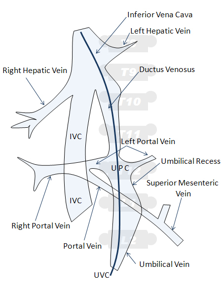
- Goal is to place the UVC above the diaphragm but below the heart
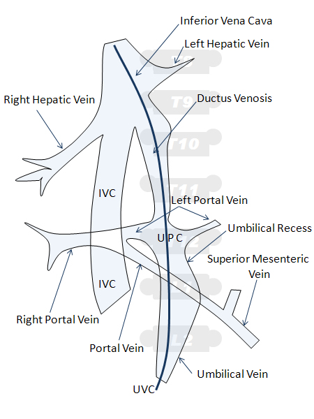
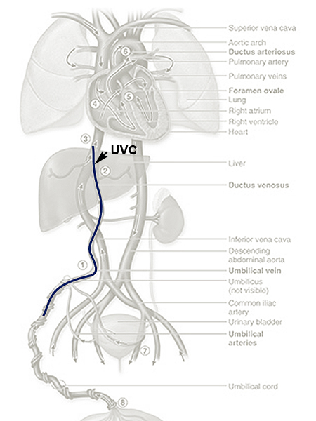
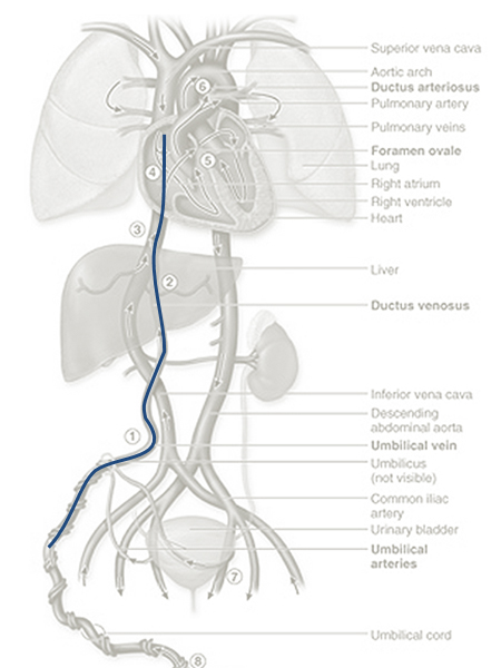
- UVC is directed into the umbilical vein, through the ductus venous, into the inferior vena cava
- Allows infusion of more hypertonic solutions
- Allows better nutrition through TPN
- Provides emergency access for fluids, resuscitation drugs and blood products
<img align="bottom" alt="Umbilical catheter position" height="600" src="http://image.wikifoundry.com/image/1/GWJuIjLId0OcSBW_Q3X4rg131678/GW450H600" title="Umbilical catheter position" width="450"/>
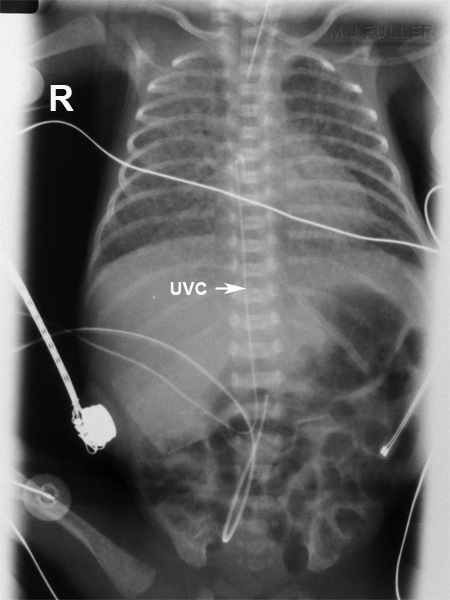
adapted from source: unknownThe umbilical vein catheter (UVC) takes a characteristically different path to the Umbilical Artery Catheter (UAC).
See the lateral decubitus abdominal image below for further appreciation of the course of the umbilical vein.
- The umbilical vein is 2-3cm long and 4-5mm in diameter
- From the umbilicus, it passes cephalad and a little to the right It joins the left branch of the portal vein after giving off several large intrahepatic branches.
- The ductus venosus arises from the point where the UV joins the left portal vein
UVC
This lateral image gives an appreciation of the course of the umbilical vein and ductus venosis in relation to the liver
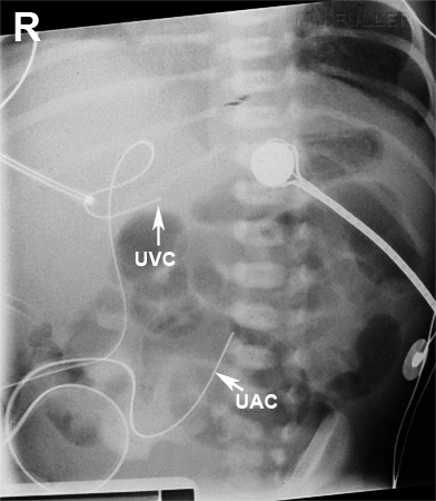
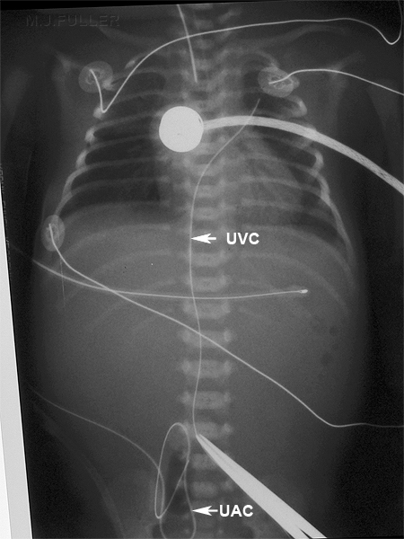
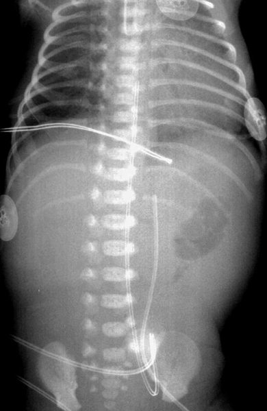
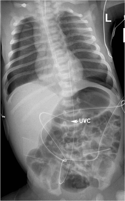
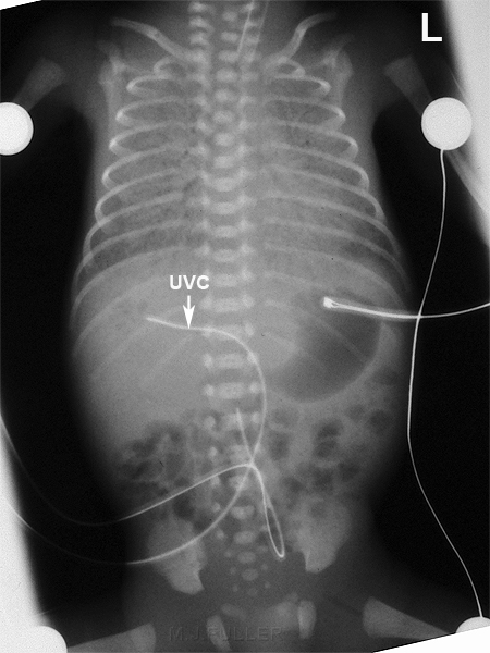
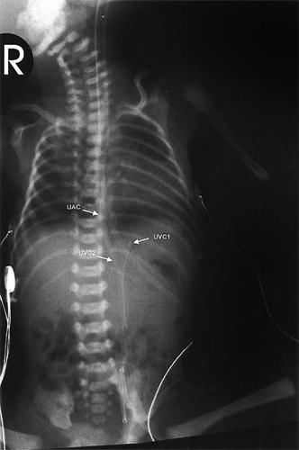
<a class="external" href="http://www.hawaii.edu/medicine/pediatrics/neoxray/neoxray.html" rel="nofollow" target="_blank">http://www.hawaii.edu/medicine/pediatrics/neoxray/neoxray.html</a>In this image the UAC is on the baby's right and the UVC is on the baby's left. This is the reverse of the usual configuration. Can you explain this anomaly?
hint1: scroll up the page and look at the lines in the lateral supine decubitus abdominal image
hint 2: look at the ribs and pelvis
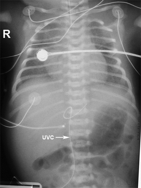
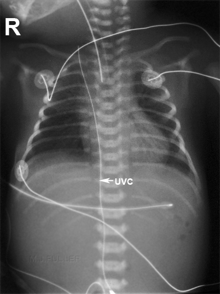
Original artwork by M.J.FullerAgain, the UVC may have looped at the junction of the left portal vein and then travelled up the left portal vein
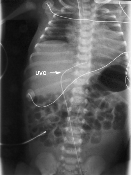
Original artwork by M.J.FullerThe UVC has deviated to the left at T8 which is the level of the left hepatic vein. The UVC tip is most likely to lie within the left hepatic vein
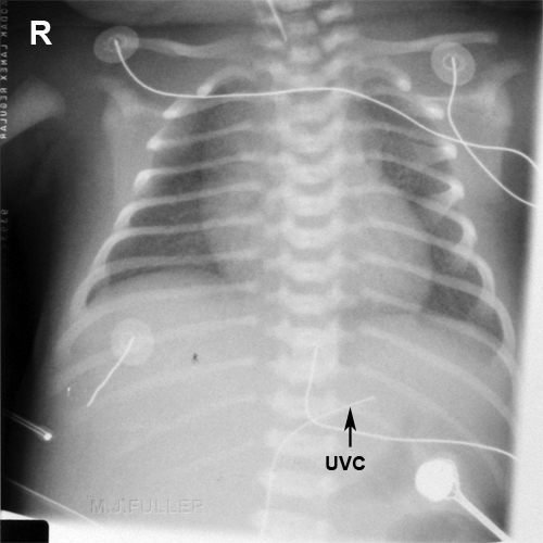
Original artwork by M.J.FullerA similar appearance to the case above but at T11. This is the level of the left portal vein and most likely represents its tip position.
<a class="external" href="http://www.youthclinic.com/Pediatric+lectures/Line+Placement.ppt" rel="nofollow" target="_blank">www.youthclinic.com/Pediatric%20lectures/Line%20Placement.ppt</a>
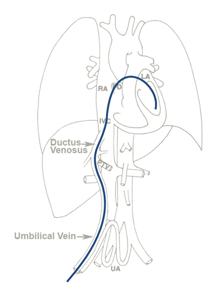
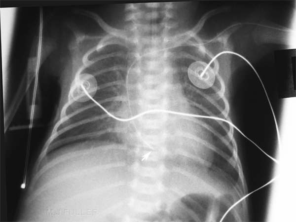
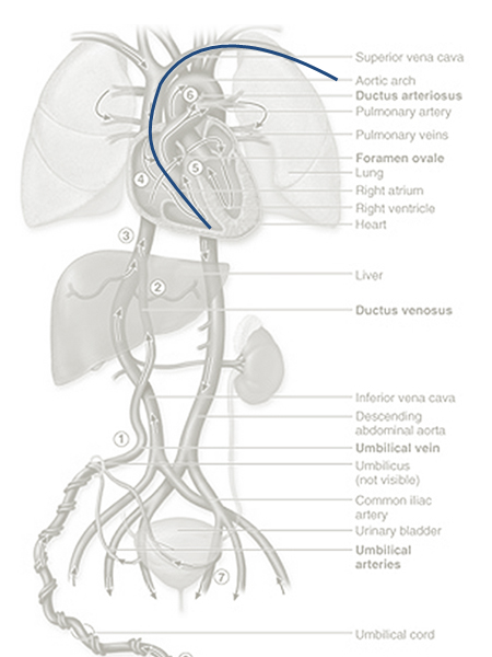
<a class="external" href="http://radiographics.rsnajnls.org/cgi/reprint/11/5/849?maxtoshow=&HITS=10&hits=10&RESULTFORMAT=&fulltext=umbilical+vein&searchid=1&FIRSTINDEX=0&sortspec=relevance&resourcetype=HWCIT" rel="nofollow" target="_blank">adpated from Lakshmana Das Narla, MD Mark Horn, MD GaryK Lofland, MD Williarn B. Moskowitz, MD. Evaluation of Umbilical Catheter and Tube Placements in Premature Infants</a>UVC extends across foramen ovale into the left atrium then left ventricle UVC extends across foramen ovale into the left atrium then left ventricle
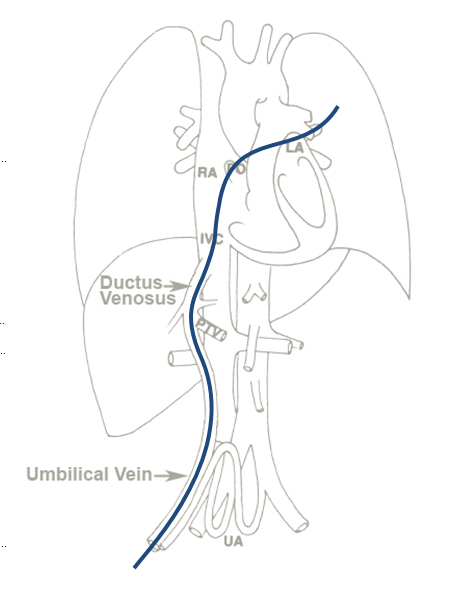
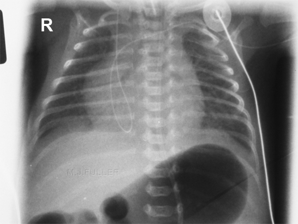
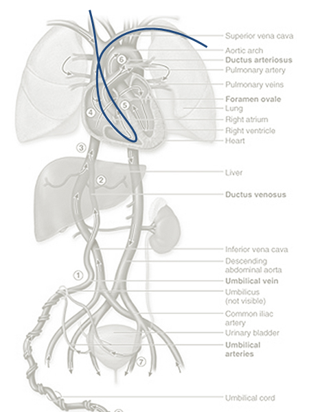
<a class="external" href="http://radiographics.rsnajnls.org/cgi/reprint/11/5/849?maxtoshow=&HITS=10&hits=10&RESULTFORMAT=&fulltext=umbilical+vein&searchid=1&FIRSTINDEX=0&sortspec=relevance&resourcetype=HWCIT" rel="nofollow" target="_blank">adpated from Lakshmana Das Narla, MD Mark Horn, MD GaryK Lofland, MD Williarn B. Moskowitz, MD. Evaluation of Umbilical Catheter and Tube Placements in Premature Infants</a>The UVC tip is in the pulmonary vein UVC through foramen ovale into the left atrium and pulmonary vein. The Foramen Ovale is a hole between the right and left atria allowing blood to pass from the right side of the heart to the left without passing through the lungs.
adapted from source: unknownSame patient as above
UVC repositioned- now in jugular vein
<a class="external" href="http://www.youthclinic.com/Pediatric+lectures/Line+Placement.ppt" rel="nofollow" target="_blank"></a>
adapted from source: unknownUVC well into jugular vein
? UVC in UA (double lumen catheter)and UAC in UV (single lumen catheter
adapted from source: unknownUVC tip in right atrium UVC tip in right atrium
Original artwork by M.J.FullerThe UVC deviates to the right at the level of the right portal vein. ? Air in portal venous branches associated with umbilical venous catheter insertion
Transient Portalvenous air
The UVC tip has tracked into the liver and there appears to be portal venous gas.
Air in portal venous branches can be associated with umbilical venous catheter insertion. Inconsequential transient portal venous air can be seen immediately after umbilical venous catheter insertion and should not necessarily be attributed to necrotizing enterocolitis.
<a class="external" href="http://www.ajronline.org/cgi/content/full/180/4/1147" rel="nofollow" target="_blank">Alan E. Schlesinger1, Richard M. Braverman1 and Michael A. DiPietro2 </a><a class="external" href="http://www.ajronline.org/cgi/content/full/180/4/1147" rel="nofollow" target="_blank">AJR 2003; 180:1147-1153</a>
Double Catheter Technique
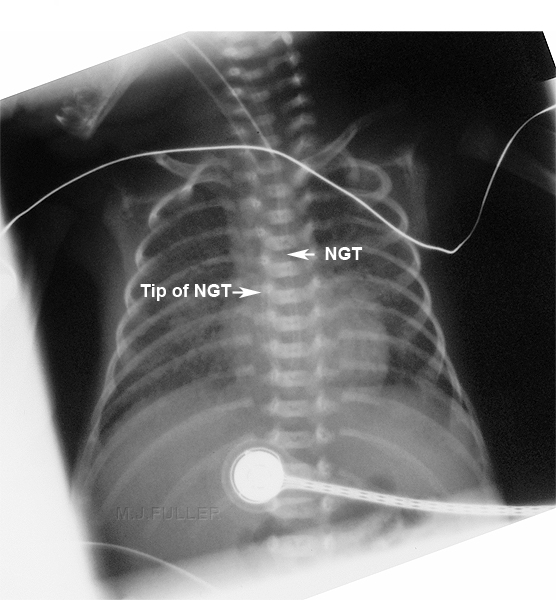
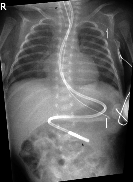
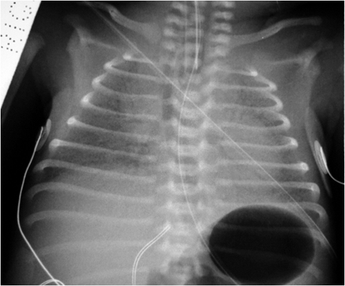
Nasogastric Tube
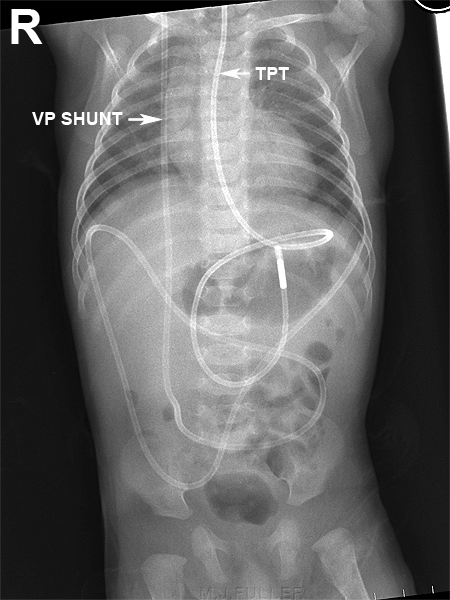
Transpyloric Tube and VP Shunt
Peripheral Venous Catheter (Long Lines, PICC, CVC)
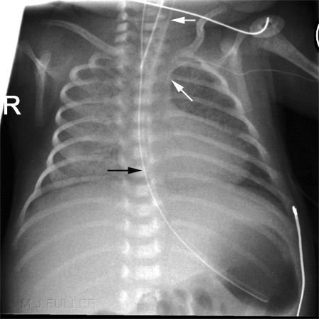
Starting at the top:
- endotracheal tube (black outline arrow)
- peripherally inserted venous line (white outline arrow)
- Nasogastric tube (solid white arrow)
- Transpyloric tube (solid black wrrow)
Peripheral Venous Catheter Insertion Sites
"1.Hand
Dorsal arch veins
Are best seen on the back of the hand but are usually larger and easier to enter just over the back of the wrist. Skin entry should be more distally. These veins can often be palpated in a larger infant rather better than they can be seen. IV's inserted here are easily splinted and any infiltration easily spotted, so are the preferred site.
Cephalic Vein, in anatomical snuffbox
This is often quite large, especially if the dorsal arch is well used. This vein can often be felt rather better than it can be seen and is one of the veins to try if one must do it "blind" in a large baby. The feeder veins over the dorsum of the hand in the first interspace need to be treated with respect, as it is possible to cannulate an artery here, risking loss of a thumb, or part thereof. (princeps pollicis artery). This is present in about 10% of infants. If present is usually the sole supply to thumb.
The cephalic vein is large and tends to last quite well. It is a good secondary site. It can also be used for insertion of percutaneous central venous catheters.2.Wrist
Volar aspect
Veins are easily seen on the volar side of the wrist. They are usually quite small and fragile and, while easily cannulated, do not last well.
They are useful secondary sites, but must be carefully watched when noxious substances (eg Dopamine, Vancomycin) are infused, as they are prone to "burn".3.Cubital Fossa
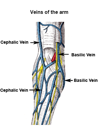
Median antecubital, Cephalic and Basilic veins
Are easy to hit and tend to last quite well if splinted properly. Median nerve and brachial artery are both vulnerable. These veins are the preferred sites for insertion of percutaneous central venous catheters. These should be avoided unless absolutely necessary in any infant likely to need long term IV therapy.
4.Foot
Dorsal arch
Are small, but easily cannulated and last surprisingly well. Vein on lateral aspect, running below malleolus, is easy of access but must be splinted carefully and watched for infiltration.
Veins leading up to short saphenous are often good value.Saphenous vein, ankle
Runs reliably just anterior to medical malleolus and is large and straight. Easy to access and lasts well although not always readily visualised.
5.Leg
Saphenous vein. Knee
Runs just behind medial aspect of knee. Often visible both behind knee and as it curves around top of tibia. Access is easy and lasts well if properly splinted. However, this vein is a good site for the insertion of percutaneous central venous catheters and should be avoided if possible in any infant likely to need long term IV therapy.
6.Scalp
Superficial temporal
Runs anterior to ear and is accessible over a distance of 5-8 cm. in most babies. Is accessible and lasts well. However, this vein is a good site for the insertion of percutaneous central venous catheters and should be avoided if possible in any infant likely to need long term IV therapy.
One hazard is the proximity of the temporal artery, which runs beside it. In small infants it can be almost impossible to tell the difference, even when the catheter has been inserted. It is important to try to identify the vessels separately, by careful palpation and by observation in a good light (in the smaller infants one can see the artery pulsate). If the catheter is in an artery, it must be removed.Posterior auricular
A moderate sized vein runs behind the ear and over the temporal bone. This vein and its branches are accessible and last quite well. There is a moderate risk of cannulating an artery in this area as well, so care must be taken.
Supratrochlear vein
This vein runs down from scalp over forehead and bridge of nose. It usually needs a shave of the hair to be accessible. Is quite easy to access and easily secured and lasts well.
The major drawback is that that the vein has anastomoses via the orbital plexus with the cavernous sinus. Thrombophlebitis of the vein has therefore a direct communication intracranially.
A further disadvantage is that any 'burn' caused by infiltration will be in the middle of the forehead. Because of these factors, it should only be used as a vein of last resort.
Scalp veins should only be used once other alternatives are exhausted. Most involve at least partial shaving of the head. Infants will take 6-12 months to grow hair back properly. This distresses parents and makes the baby look 'funny' for the first year or so."<a class="external" href="http://www.rch.org.au/nets/handbook/index.cfm?doc_id=11235" rel="nofollow" target="_blank">Source Neonatal Handbook, Royal Childrens Hospital, Melbourne
</a>
Radiography Technique
Upper Limb
Check your departmental policy regarding the arm position for venous catheters.
The Southern West Midlands Newborn Network recommend the following
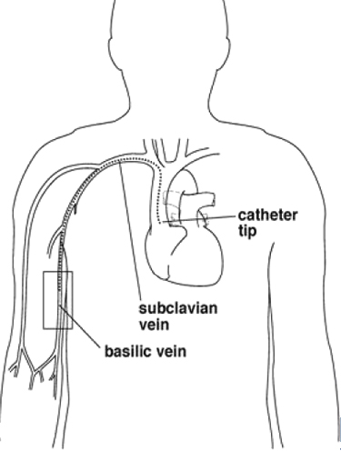
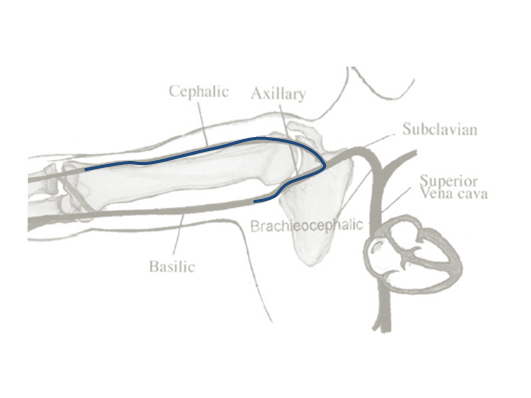
It is important to note that the catheter tip position will change with the baby's arm movements. The following research finding are noteworthy"If the cephalic vein has been cannulated the x-ray should be taken with the arm adducted. If the basilic vein has been cannulated the x-ray should be taken with the arm abducted.
Contrast medium may be used to aid visualisation of the line.
Although not reported in neonates, there is a theoretical risk of reaction to the contrast medium.
The tip of the PICC should lie outside the heart, ideally by 1cm in a baby <1250g and by 2cm in a baby >1250g."
<a class="external" href="http://www.newbornnetworks.org.uk/southern/PDFs/InsertionofCVLfinalv1Dec05.pdf" rel="nofollow" target="_blank">(</a><a class="external" href="http://www.newbornnetworks.org.uk/southern/PDFs/InsertionofCVLfinalv1Dec05.pdf" rel="nofollow" target="_blank">Southern West MIidlands Newborn </a><a class="external" href="http://www.newbornnetworks.org.uk/southern/PDFs/InsertionofCVLfinalv1Dec05.pdf" rel="nofollow" target="_blank">Network)</a>
"Arm movements were associated with significant
displacement of catheters. Catheters that were
placed via the basilic or axillary vein migrated toward the
heart with adduction of the arm, whereas those that were
placed via the cephalic vein moved away from the heart
with adduction. Flexion of the elbow displaced catheters
that were placed in the basilic or cephalic vein below the
elbow toward the heart but did not have any effect on
catheters that were placed via the axillary vein. For catheters
that were placed in the basilic vein, simultaneous
shoulder adduction and elbow flexion caused the greatest
movement toward the heart (15.11 + or - 1.22 mm). We
were able to reposition correctly inappropriately placed
catheters in 9 of 10 patients by using arm movements."<a class="external" href="http://pediatrics.aappublications.org/cgi/reprint/110/1/131" rel="nofollow" target="_blank">Ali M. Nadroo, Ronald B. Glass, Jing Lin, Robert S. Green and Ian R. Holzman</a>
<a class="external" href="http://pediatrics.aappublications.org/cgi/reprint/110/1/131" rel="nofollow" target="_blank">Central Catheters in Neonates, Pediatrics 2002;110;131-136</a>
The person who has inserted the catheter may not appreciate it being repositioned by the radiographer!
source: unknown
Source <a class="external" href="http://pediatrics.aappublications.org/cgi/reprint/110/1/131" rel="nofollow" target="_blank">Ali M. Nadroo, Ronald B. Glass, Jing Lin, Robert S. Green and Ian R. Holzman</a><a class="external" href="http://pediatrics.aappublications.org/cgi/reprint/110/1/131" rel="nofollow" target="_blank"> Central Catheters in Neonates, Pediatrics 2002;110;131-136</a>
Source <a class="external" href="http://pediatrics.aappublications.org/cgi/reprint/110/1/131" rel="nofollow" target="_blank">Ali M. Nadroo, Ronald B. Glass, Jing Lin, Robert S. Green and Ian R. Holzman</a><a class="external" href="http://pediatrics.aappublications.org/cgi/reprint/110/1/131" rel="nofollow" target="_blank"> Central Catheters in Neonates, Pediatrics 2002;110;131-136</a>Note the position of this CVC in a baby with arm abducted and elbow extended Same baby with same CVC line with arm adducted and elbow flexed.
Central Venous Catheter (CVC) Longline- leg
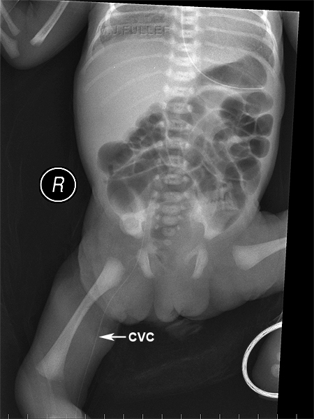
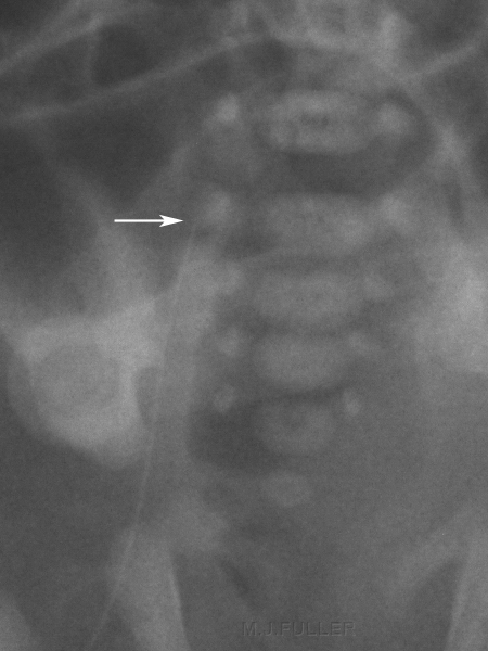
The central venous catheter has been inserted in the right leg and its tip is difficult to localise precisely. This is a common problem with CVC lines and emphasises the need for good quality images when checking CVC lines. A study of printed CR images vs soft copy CR images reported a long line detection rate of 66.7% vs 95.6% respectively. <a class="external" href="http://www.pubmedcentral.nih.gov/picrender.fcgi?artid=1721656&blobtype=pdf" rel="nofollow" target="_blank">(A Evans, J Natarajan, C J Davies, 2004).</a>
Other suggestions include
"The authors emphasise the importance of verifying neonatal long line position using contrast, as the exact localisation of the catheter tip can be difficult on plain radiographs. As an alternative, Groves et al4 have described the use of colour Doppler to aid ultrasonographic line tip visualisation." <a class="external" href="http://fn.bmj.com/cgi/content/extract/91/4/F311" rel="nofollow" target="_blank">T M Berger, M Stocker, J Caduff</a>
Gonad protection could have been used to good effectA magnified view demonstrates the CVC tip to be at the level of the first sacral vertebra
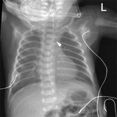
Central Venous Catheter (PICC) - arm
source: unknown
adapted from source: unknownContrast injected through a subclavian line. ? tip in jugular vein
Injection of contrast into jugular CV line demonstrates filling of the vascular plexus in the right shoulder as well as filling of the subclavian vein and the axillary vein
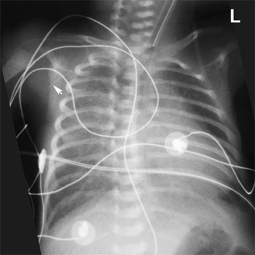
adapted from source: unknownCentral line in Right Ventricle (arrow)
adapted from source: unknownLong left central line in right heart then subclavian vein
adapted from
<a class="external" href="http://pediatrics.aappublications.org/cgi/reprint/110/1/131" rel="nofollow" target="_blank">Ali M. Nadroo, Ronald B. Glass, Jing Lin, Robert S. Green and Ian R. Holzman
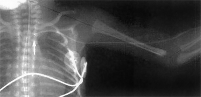
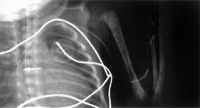
Central Catheters in Neonates, Pediatrics 2002;110;131-136</a>Central line inserted in cephalic vein with tip in right axillary vein (tip arrowed) Central line in right axillary vein graphic
Intercostal Drain
Endotracheal Tube
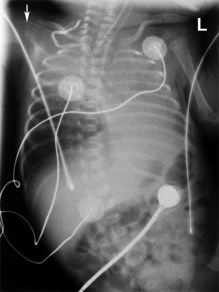
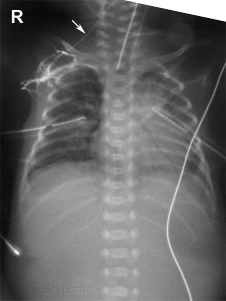
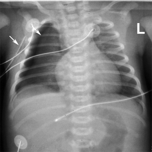
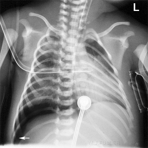
This baby has a right sided pneumothorax. The top white arrow identifies the lung edge
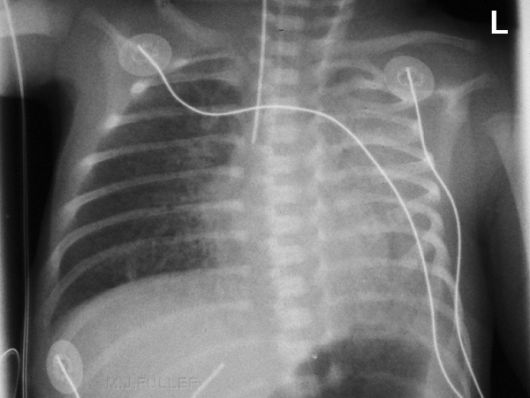
An intercostal drain is malpositioned in the soft tissues of the chest wall.
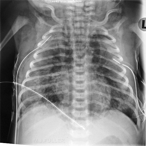
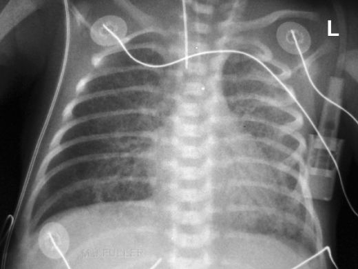
This baby has extensive subcutaneous emphysema. Both intercostal drains are malpositioned with their sideholes possibly in the chest wall soft tissues
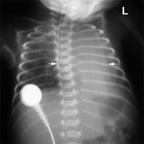
This baby has a right sided pneumothorax. Also note
- LPO position
- intercostal tube malposition
- mediastinal shift
- deep suclus sign (white arrow)
- silhouette sign right hemidiaphragm
- NGT
ET tube placement, should be halfway between the thoracic inlet and the carina
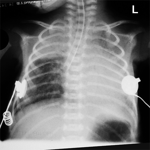
The endotracheal tube tip is in the right main bronchus. A hint of opacity in the RUL suggests that it may be partly within the bronchus intermedius. There is atelectasis of the left lung. There are several skin folds over the left lung
- About 10% of ETT are initially placed in the right main stem bronchus
- With time, the left lung becomes atelectatic
- If on ventilator, the right lung may be hyperinflated
http://www.learningradiology.com/archives04/COW%20129-Atelectasis-ETT/atelectasiscorrect.htm
- May lead to pneumothorax or tension pneumothorax
Point of Interest
</a>
The endotracheal tube (ETT) tip is in the bronchus intermedius.
When the tip is in the bronchus intermedius, RUL will also become atelectatic along with all of left lung
This baby has an ET tube malpositioned in the oesophagus. Note the baby's trachea has deviated to the right and is positioned alongside the ET tube
...back to the Applied Radiography home page
