Neck of Humerus Fractures

From wikiRadiography
Jump to navigation Jump to search
Introduction

Neck of humerus fractures are commonly seen in the Emergency Department particularly in elderly patients following a fall. This page considers all aspects of radiography of neck of humerus fractures in all age-groups. The term neck of humerus fracture is interpreted broadly to include all proximal humerus fractures.


Anatomy
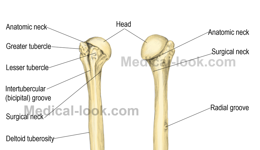
proximal humerus anatomy
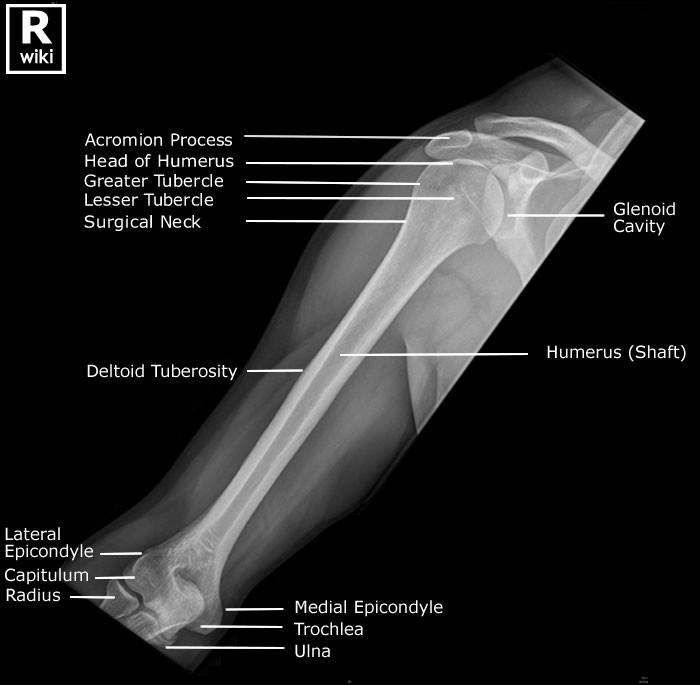
Radiographic Anatomy - Humerus AP


Pseudosubluxation and Lipohaemarthrosis
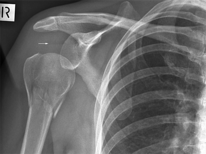
NOH fracture
This patient has fractures of the neck of humerus and greater tuberosity. The humeral head is subluxed inferiorly and this subluxation is likely to be caused by distension of the joint by joint effusion (haemarthrosis). There is a fat/fluid level consistent with lipohaemarthrosis of the shoulder joint (white arrow). This indicates the presence of intraarticular injury. A lipohaemarthrosis will only be seen when patients are imaged with a horizontal beam (i.e. erect).


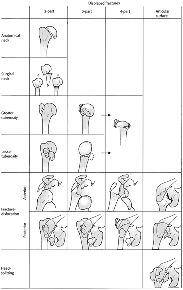
The Neer's Classification of Proximal Humerus Fractures

Neers Classification Proximal Humerus Fractures
<a class="external" href="http://www.msdlatinamerica.com/ebooks/PracticalOrthopaedicSportsMedicineArthrocopy/sid280790.html" rel="nofollow" target="_blank">http://www.msdlatinamerica.com/ebooks/PracticalOrthopaedicSportsMedicineArthrocopy/sid280790.html</a>


Radiographic Technique

Transthoracic Lateral

transthoracic lateral
The transthoracic lateral projection of the shoulder/proximal humerus is a technique that is not commonly practiced by most radiographers. The technique is a valid method for radiographic demonstration of the proximal humerus and is worthy of inclusion in the radiographers' technique toolkit.

The reasons for the lack of favour of this technique are likely to be
  • the inherent superimposition of the anatomy over the patient's chest
  • the need for the patient to hold very still during a relatively long exposure
  • and the fact that it is a very operator-dependent technique.. it takes some practice to master

Further information here


Case 1

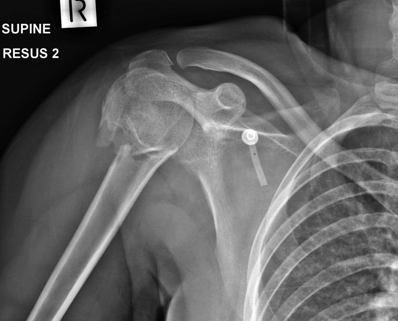
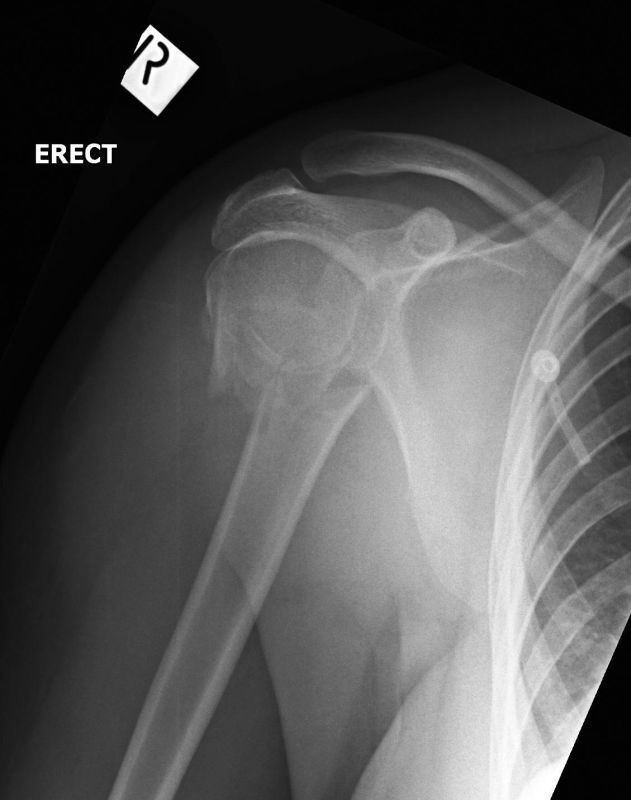
AP shoulderThis 25 year old female presented to the Emergency Department following a fall from a pushbike. She was assessed in the resus room and radiography including her right shoulder was requested.

AP shoulder radiography was performed with a wireless DR-X system using a bucky tray.There is a fracture of the surgical neck of humerus.
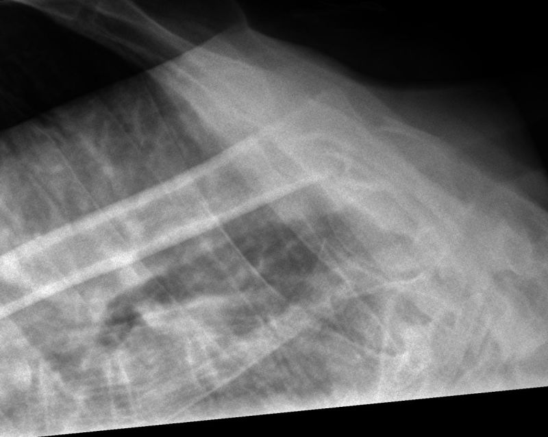
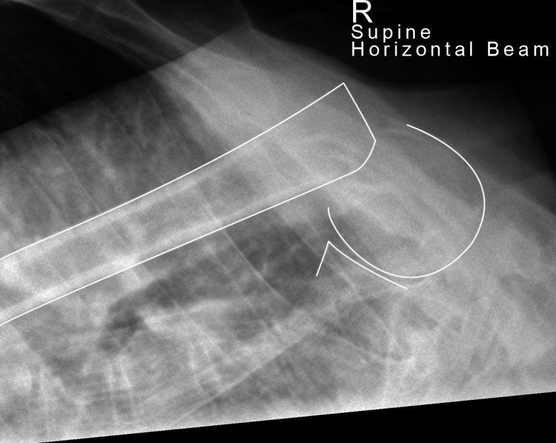
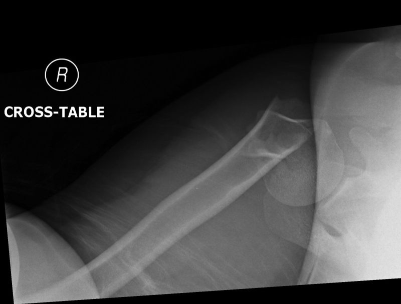
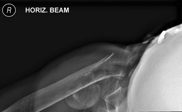
transthoracic lateral shoulderThe patient could not easily be positioned for lateral scapula or IS shoulder radiography. The radiographer performed a transthoracic lateral view of the right shoulder to demonstrate alignment of the fracture. This technique does not demonstrate detailed bony anatomy but is valuable in demonstrating the true alignment/position of the fracture in a true lateral position.

A DRX-1 wireless Digital X-ray Imaging System was utilised without stationary grid. A long exposure time technique was employed.
transthoracic lateral shoulderConsiderable foreshortening and angulation at the fracture site was demonstrated in the transthoracic lateral projection.
IS shoulderAn IS projection was considered worthwhile in terms of demonstrating bony detail.
transthoracic lateral shoulderProximal humerus fractures can be treated conservatively with a collar-and-cuff or operatively with ORIF. The decision will be influenced by the radiographic assessment of the position of the fracture fragments. The supine transthoracic lateral does not give a true representation of the position that the fracture is likely to heal in- the reason for this is that the collar-and-cuff treatment uses the weight of the patient's arm as a method of reducing the fracture with the patient in the erect position. What is required is a radiographic demonstration of the fracture in the position that it would heal in if treated conservatively.

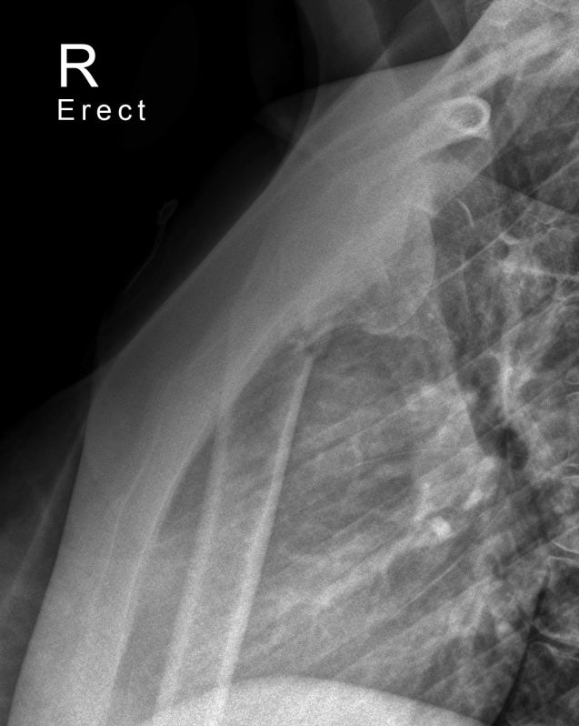
Following further assessment of the patient's injuries, it was considered safe for the patient to be positioned in an erect sitting position. The transthoracic lateral projection was repeated with the patient in the erect position.

Whilst the technique has limitations, the fracture is shown with improved alignment in the erect position when compared to the supine position above.
AP shoulderThe erect AP shoulder showed no improvement in the alignment of the neck of humerus fracture.


Case 2
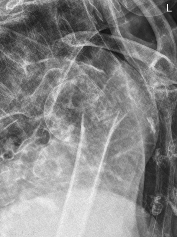
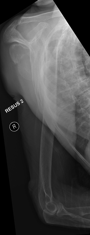
Neck of Humerus Fractures - wikiRadiographyNeck of Humerus Fractures - wikiRadiographyThis 85 year old lady presented to the Emergency Department after falling onto her right shoulder. She was examined and referred for right shoulder radiography.

The radiographer has exercised caution by including the entire right humerus. There is a comminuted fracture of the anatomical neck of humerus.

This is a greater tuberosity 3 part fracture using the Neer's classification of neck of humerus fractures (see above)
Neck of Humerus Fractures - wikiRadiographyThe radiographer also performed an IS projection of the right shoulder which further demonstrated the degree of displacement of the head of humerus.

The patient subsequently underwent shoulder replacement surgery.


... back to the Wikiradiography home page
... back to the Applied Radiography home page