Multi-lobar Collapse
Jump to navigation
Jump to search
Introduction
Combined RML and RLL collapse
... back to the Applied Radiography home page
Multilobar collapse can produce confusing patterns on chest X-ray images. This page considers patterns of multilobar collapse.
Combined RML and RLL collapse
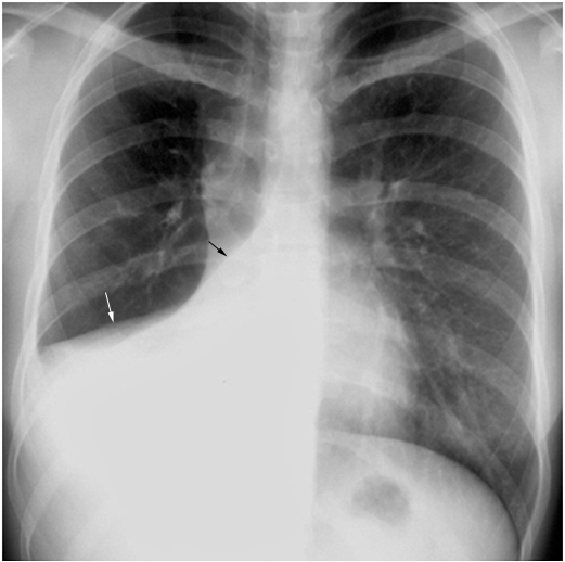
source: Paul Stark (MD)This uncommon combination can produce a confusing plain film appearance. The cause of the collapse often involves tumour compression of the bronchus intermedius. Loss of visualisation of the right hemidiaphragm and right heart border requires consideration of a combined RML and RLL collapse.
There is loss of visualisation of the right hemidiaphragm and right heart border suggestion RLL and RML disease respectively.
The oblique fissure is demonstrated in a collapsed position (black arrow) and the horizontal fissure as similarly seen in a collapsed position (white arrow)
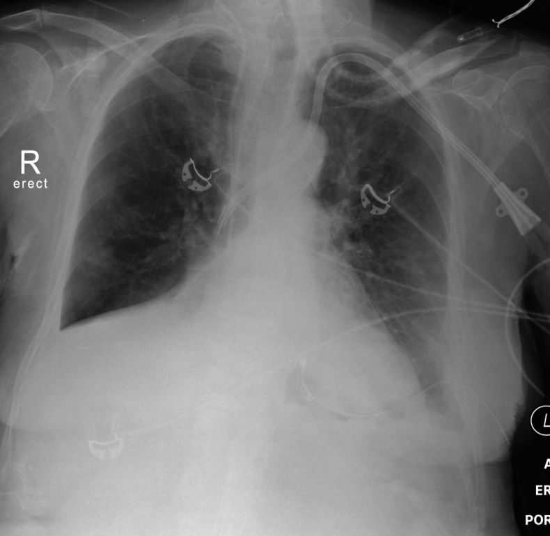
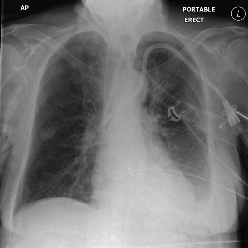
Case 1
... back to the Applied Radiography home page


