Monteggia and Galeazzi Fracture-dislocations of the Forearm
The Monteggia and Galeazzi are unstable fracture-dislocations of the forearm. The Monteggia fracture-dislocation features a dislocation of the radius at the elbow and the Galeazzi fracture-dislocation involves a dislocation of the ulna at the wrist. An awareness of these injuries can assist the radiographer to demonstrate them adequately.
The Monteggia Fracture
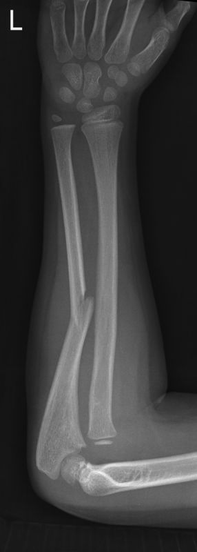
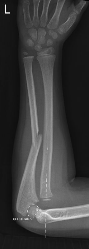
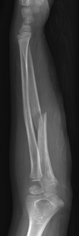
Case Study 1.
This 7 year old girl presented to the Emergency Department after falling onto her left arm. She was examined and referred for left forearm radiography.
The Galeazzi Fracture
History
<a name="IntroductionHistoryoftheProcedure"> </a>The Galeazzi fracture injury pattern was first described 1842, by Cooper, 92 years before Galeazzi reported his results. Ricardo Galeazzi (1866-1952), an Italian surgeon at the Instituto de Rachitici in Milan, was known for his extensive work experience on congenital dislocation of the hip. In 1934, he reported on his experience with 18 fractures with the above-described pattern as a compliment to the Monteggia lesion. Such fractures have since become synonymous with his name. (<a class="external" href="http://emedicine.medscape.com/article/1239331-overview" rel="nofollow" target="_blank">source: emedicine</a>)
Case Study 1.
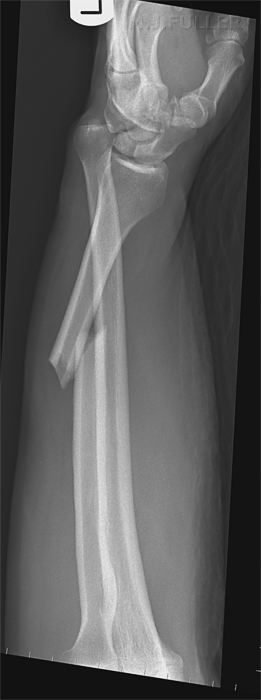
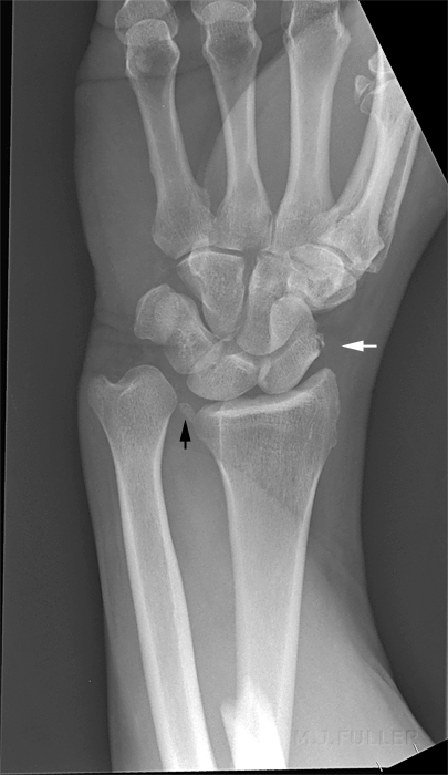
This patient presented following a fall from his pushbike. The shape of his forearm was not normal. The swelling from the radial fracture was evident, as was the prominence of the ulnar styloid and the radial deviation. It was clear that the patient had sustained a bony injury.
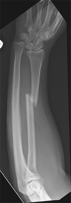
Case Study 2
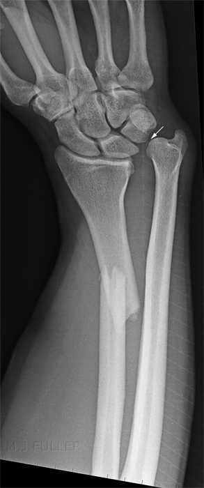
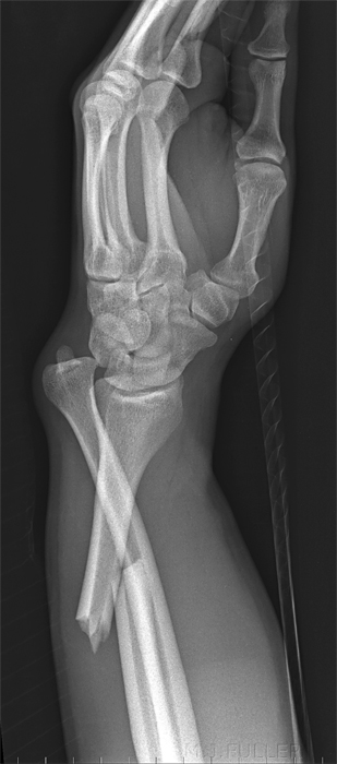
This 40 year old male presented to the Emergency Department following a fall onto an outstretched hand. He had an obvious forearm deformity.
Radiography
Patients with Galeazzi fractures will be in considerable pain, even following the administration of pain relief. If you consider the radius and ulna to form a ring of bone, the ring has been disrupted in two places and is therefore unstable. It is reasonable to perform the PA wrist projection first- this is the easiest view and, if viewed immediately, will provide a diagnosis. The lateral view is the difficult one. You could roll the patient's arm into the lateral position but this will have two disadvantages. Firstly, you may have difficulty achieving a true lateral demonstration of the wrist. Secondly, it will be very painful for the patient. An arguably better approach would be to raise the patient's forearm and support it on a positioning sponge, then perform a horizontal ray lateral view. This is the technique that was utilised in the case above. Note that the wrist is not true AP or true lateral in both views. This was noted at the time of the examination and could possibly have been corrected with tube/IR angulation.
Ulnar Variance
In cases of Galeazzi fractures, ulnar plus variance (radial shortening) of 10 mm or more implies complete disruption of the interosseous membrane and, therefore, complete instability of the distal radioulnar joint following reduction.
<a class="external" href="http://radiology.rsnajnls.org/cgi/reprint/184/1/15.pdf" rel="nofollow" target="_blank">F. A. Mann, MD . Anthony J. Wilson, MB, ChB Louis A. Gilula, MD. </a>
<a class="external" href="http://radiology.rsnajnls.org/cgi/reprint/184/1/15.pdf" rel="nofollow" target="_blank"> Radiographic Evaluation ofthe Wrist:</a>
<a class="external" href="http://radiology.rsnajnls.org/cgi/reprint/184/1/15.pdf" rel="nofollow" target="_blank"> What Does the Hand Surgeon Want to Know?’</a>
<a class="external" href="http://radiology.rsnajnls.org/cgi/reprint/184/1/15.pdf" rel="nofollow" target="_blank"> Radiology, Volume 184, Number 1</a>
Treatment
In 1941, Campbell termed the Galeazzi fracture the "fracture of necessity, "because it necessitates surgical treatment in adults- nonsurgical treatment of the injury results in persistent or recurrent dislocations of the distal ulna.(<a class="external" href="http://emedicine.medscape.com/article/1239331-overview" rel="nofollow" target="_blank">source: emedicine</a>)
Among 33 patients with a Galeazzi-type fracture-dislocation of the forearm, there were two children and 26 adults with a classic Galeazzi injury, and five patients with a Galeazzi-equivalent lesion. The worst results were obtained in type-I lesions. Closed reduction was primarily successful in children. The results of surgical treatment were much better in adults. It is advisable to treat this complex injury by anatomic reduction and internal fixation of the radial shaft fracture. Immobilization in a fully supinated position is recommended to reduce the dislocation of the distal radioulnar joint. Additional temporary radio-ulnar fixation with Kirschner wires is also necessary in cases of severe derangement of the distal radioulnar joint.
<a class="external" href="http://www.ncbi.nlm.nih.gov/pubmed/8145315?dopt=Abstract" rel="nofollow" target="_blank">Maculé Beneyto F, Treatment of Galeazzi fracture-dislocations. J Trauma. 1994 Mar;36(3):352-5.</a>
OtherIn Galeazzi fractures, the interosseous membrane acts as a constraint to radial shortening. Anatomic reduction with internal fixation is indicated for this fracture-dislocation. (<a class="external" href="http://www.wheelessonline.com/ortho/the_interosseous_membrane_of_the_forearm_structure_and_its_role_in" rel="nofollow" target="_blank">Schneiderman et al 1993 The Interosseous Membrane of the Forearm in Wheeless</a>)
back to the Applied Radiography home page
