Missed Fractures- Peripheral Pathology

From wikiRadiography
Jump to navigation Jump to search
Introduction

It is self-evident that a significant plain film pathology that can be demonstrated radiographically should not be overlooked. When missed plain film pathology cases are examined, they tend to fall into a few recognisable groups. This page considers cases of missed or potentially missed plain film pathologies that tend to be overlooked because they are peripherally sited on the image- that is, they are located around the edges of the image.

Case 1
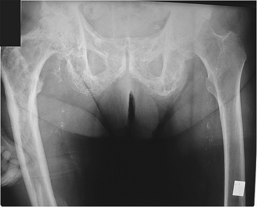
pelvis This patient has presented following a fall. The provisional diagnosis is fractured right neck of femur. Can you see a fractured neck of femur?


Discussion
Your eye is naturally focused on the right neck of femur. Distractions include evidence of Paget's disease, an ID window that is partially obscuring the right femur and a few phalanges overlying the right femur. What is important in this image is the anatomy at the bottom of the image- the patient has a right midshaft femur fracture.

Interestingly, the patient's hand is sometimes a clue as to the location of the pathology- patients tend to grab/support their painful anatomy


Case 2
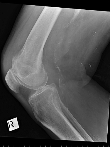
KNEE This patient also presented following a fall and was referred for knee radiography. There is evidence of a small knee effusion and metal sutures. The examination was reported as basically normal.


see below


A deliberate gap here!


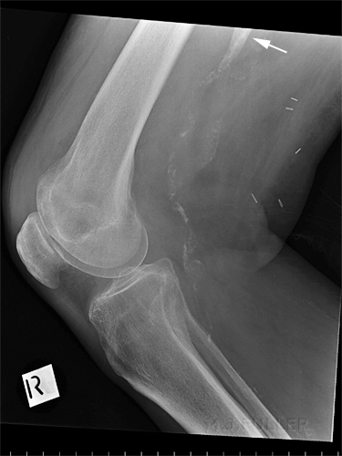
KNEEWhat was missed by all concerned was that the patient sustained a fracture of the femur. The tip of the proximal fragment is arrowed. It is unfortunately overlying the calcified femoral artery- this does not help to draw your eye to the fracture. You can closely examine the knee joint and ponder the presence of cortical breaks and soft tissue signs for a very long time and still not see the fracture.


Case 3

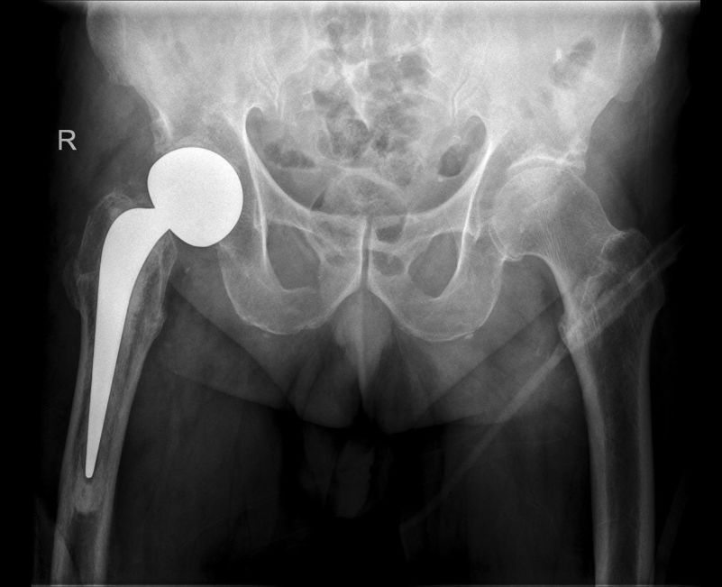
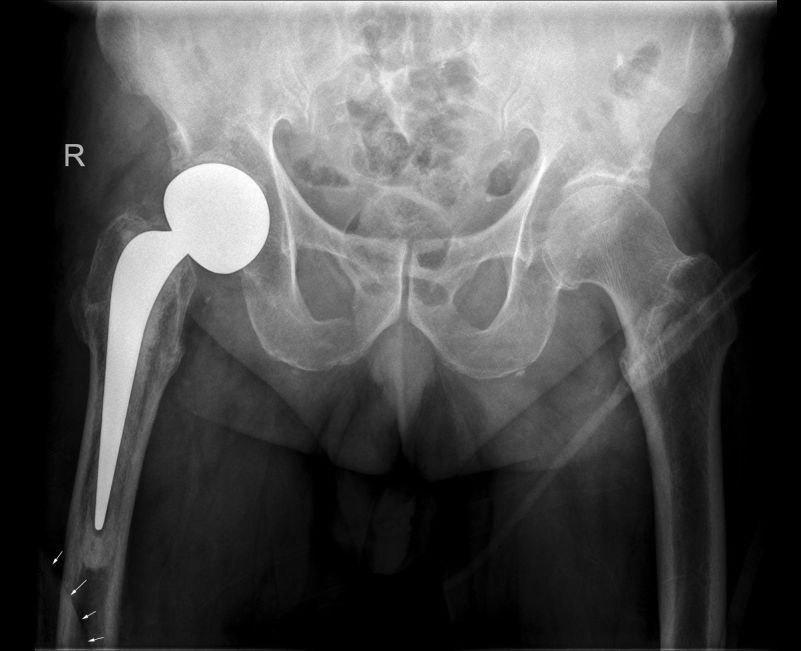
AP pelvisThis 77 year old demented male was referred to the Emergency Department after a fall onto his right hip at the nursing home. He was referred for right hip radiography. Can you see any pathology?
AP pelvisThe arrowed structure is the superior margin of the distal fragment of a spiral fracture of the femur.
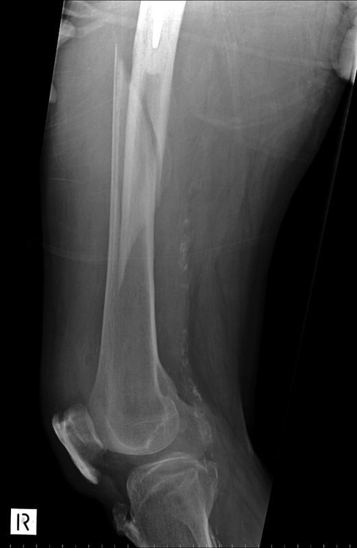
femurThe spiral fracture was unmissable on the femur projection image.

Discussion

It is good radiographic practice (in trauma) to include the entire femur when a patient is referred for pelvis/hip radiography and a hip prosthesis/screw is revealed.