Metacarpal Fractures
IncidenceMetacarpal fractures are commonplace- they account for 30 to 40 percent of all hand fractures <a class="external" href="http://www.utdol.com/patients/content/topic.do?topicKey=%7E9DlTT3y2/Um_d9" rel="nofollow" target="_blank">(Bloom J and Burrough, J. E.)</a>. This page considers all aspects of radiography of metacarpal fractures, dislocation and fracture-dislocations.
Metacarpal fractures are common. Fracture of the metacarpal bones accout for 10% of all fractures, 40% of all hand fractures: the lifetime incidence of a metacarpal fracture is 2.5%
Fractures of the 5th metacarpal make up 25% of all metacarpal fractures (which equates to 10% of all hand fractures.) [<a class="external" href="http://radiopaedia.org/articles/metacarpal-fractures" rel="nofollow" target="_blank">Dr Jeremy Jones, Radiopaedia</a>]
Base of Metacarpal Fractures/Dislocations
AnatomyCarpometacarpal dislocations occur infrequently because these joints are supported by the strength and complexity of the carpometacarpal and intermetacarpal ligaments.The second and 5th carpometaracpal joints are most commonly individually dislocated because they are more mobile than the third and fourth. Carpometacarpal fracture-dislocation most commonly involves the 5th metacarpal. The oblique view of the hand is useful in demonstrating this type of injury. (J. Harris, W. Harris and R. Novelline, The Radiology of Emergency Medicine, 3rd ed, Williams and Wilkins, 1993, p452).
"Fractures of the metacarpal base are provided a degree of stability due to the dorsal and palmar carpometacarpal ligaments as well as the interosseous ligaments. This is particularly true for 2nd and 3rd metacarpal base fractures. Fractures of the 4th, and the 5th metacarpal base, are somewhat less stable due to increased mobility at the carpometacarpal (CMC) joint. In addition, the motor branch of the ulnar nerve runs in close proximity with the 4th and 5th metacarpal bases, and accordingly, fractures of these areas warrant a thorough evaluation of ulnar-distributed motor function." <a class="external" href="http://www.utdol.com/patients/content/topic.do?topicKey=%7E9DlTT3y2/Um_d9" rel="nofollow" target="_blank">(Bloom J and Burrough, J. E.)</a>
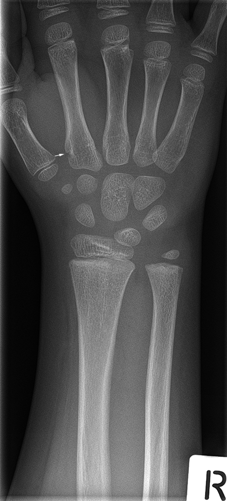
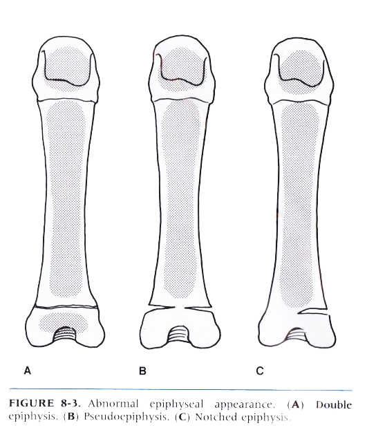
Pseudoepiphyses and Double Epiphyses
Classification of Metacarpal Fractures
Metacarpal fractures can be grouped on locations as fractures of the head, neck, shaft or base.
The Orthopaedic Trauma Association classification uses an alpha-numeric classification scheme. The schematic for metacarpal fractures is 25(modifier) - _ _ . _.
The modifier specifies which digit is involved:T - Thumb
I - Index
M - Middle
R - Ring
L - LittleThe first blank after the modifier is a letter specifying the articular nature if the fracture:
A - Extra-articular
B - Articular
C - Artcular / extraarticularThe second blank is a number specifying the bony location of the fracture:
1 - Head (includes the neck as extra-articular fractures - 25-A1)
2 - Shaft
3 - BaseThe decimal point after the first two modifiers further characterizes the fracture pattern - simple, oblique, degree of comminution, etc.
<a class="external" href="http://www.orthopaedia.com/display/Main/Metacarpal+fractures" rel="nofollow" target="_blank">Quoted from Orthopedia</a>
Case 1
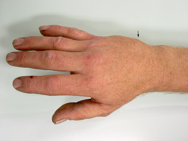
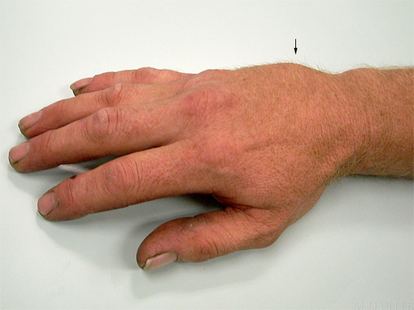
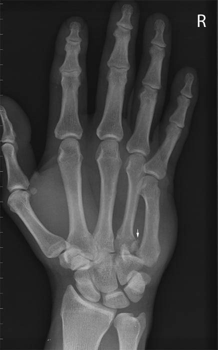
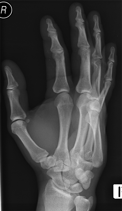
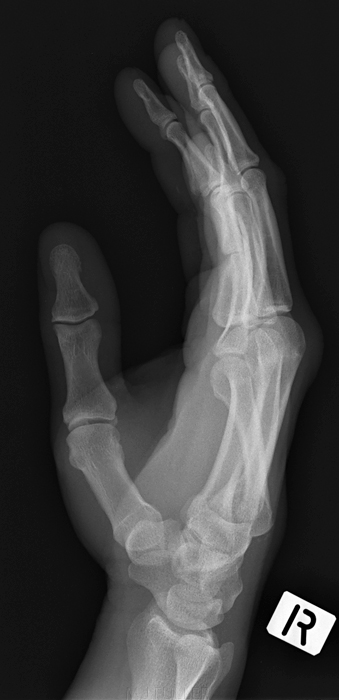
This patient presented to the Emergency Department following a fight. His right hand was very painful and he had an abnormal prominence dorsally along the ulnar aspect in the region of the base of his 5th metacarpal (arrowed). The patient's right metacarpophalangeal joint demonstrated an abnormal lack of prominence. The PA hand image demonstrates a fracture dislocation of the base of the 5th metacarpal with ulnar displacement. The oblique hand image demonstrates dorsal displacement of the dislocated 5th metacarpal. There is also a small bony fragment demonstrated at the base of the 5th metacarpal adjacent to the cortex of the hamate ? donor site The lateral hand image demonstrates the angular displacement of the 5th metacarpal base.
The fracture-dislocation was reduced with local pain relief and reimaged before the patient was taken to the operating theatre for K-wiring under image intensifier control.
Discussion"Because the fracture-dislocation of the base of the fifth metacarpal is pathologically and radiographically similar to the Bennett fracture of the thumb metacarpal, it is referred to as a"reversed Bennett", ülnar Bennett", or Tenneb"(Bennett spelled backwards) fracture". In further analogy with the Bennett Fracture, the large distal fragment of the base of the fifth metacarpal is proximally retracted by the flexor and extensor carpi ulnaris muscles, and open reduction and internal fixation are recommended to reduce and stabilise the fracture fragments. (J. Harris, W. Harris and R. Novelline, The Radiology of Emergency Medicine, 3rd ed, Williams and Wilkins, 1993, p452 - 455).
Case 2
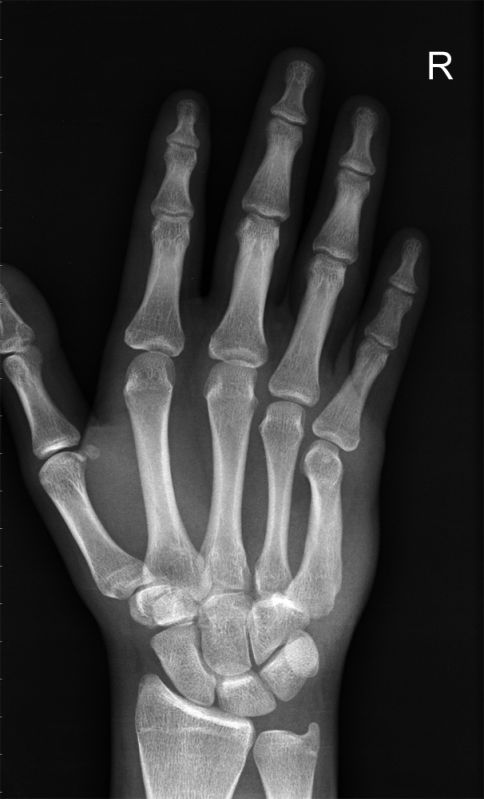
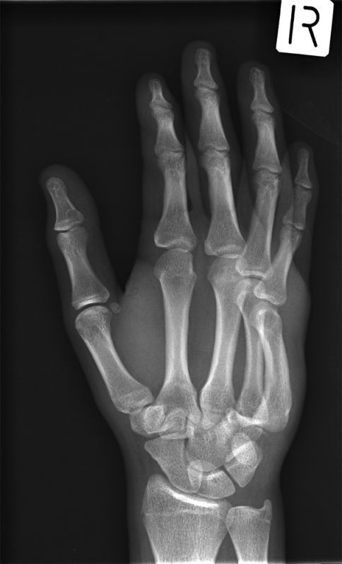
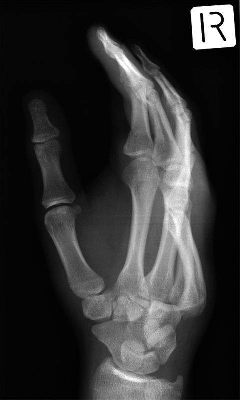
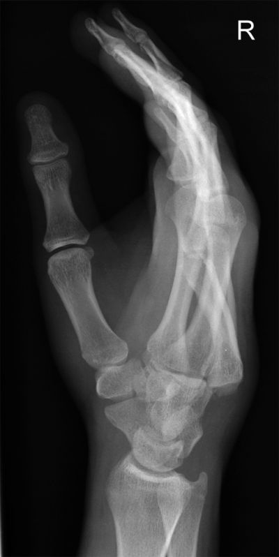
This 18 yar old male presented to the Emergency Department with a painful and swollen right hand following a fight. He had an obvious swelling over the base of his 5th metacarpal.
There is a foreshortening of the 4th and 5th metacarpal (follow the arc formed by the metacarpal heads)The oblique hand projection image demonstrates dislocation of the bases of the 4th and 5th metacarpals The hand is in a steep oblique position. There is further demonstration of posterior dislocation of the bases of the 4th and 5th metacarpals. The patient's hand is approaching lateral in this image. There 4th and 5th carpo-metacarpal dislocations are again demonstrated. There is a also a few small bony fragments at the base of the 5th metacarpal.
... back to the Wikiradiography Home page
... back to the Applied Radiography home page
