MRI Anatomy - Knee
Jump to navigation
Jump to search
This is a sagittal T1-weighted MR is through the intercondylar notch. The femur and tibia are shown as bright structures (due to the bright marrow fat signal with lots of protons) surrounded by the thin black rim of cortical bone, which has relatively few protons.
anterior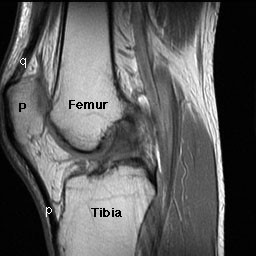 posterior
The patella (P) is seen anterior to the femur. The dark structure extending upward from the patella is the quadriceps tendon (q). The dark structure extending caudad from the patella down to the tibial tuberosity is the patellar ligament (p), sometimes called the patellar tendon. If one thinks of the patella as a bone in its own right, then the dark structure (p) coming off of its inferior tip should rightfully be called the patellar ligament, since it connects two bones. However, if you just consider the patella to be a sesamoid bone living in this really long tendon, then calling it the patellar tendon makes some sense.
Just above the patella and right behind the quadriceps tendon is the anterior suprapatellar fat pad (a). Just anterior to the femur is the prefemoral fat pad (p). Unlike the anterior suprapatellar fat pad, which is relatively constant in shape and size, this fat pad is quite variable in size, and may appear either fairly flat or quite plump. Extending up between these two fat pads is the suprapatellar bursa, an extension of the knee joint space.
anterior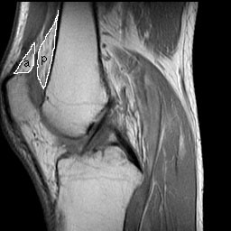 posterior
These two fat pads can usually be seen on a lateral plain film. Usually the suprapatellar bursa is only a potential space, and contains very little fluid. In this case, the two fat pads are separated by less than 5 mm. If a suprapatellar effusion is present, the two fat pads are pushed apart by the effusion. If the distance separating them is 5 mm or greater, we consider this sufficient evidence to diagnose a knee effusion on plain films. We don't use this sign with MR, because we can image the effusion directly.
The anterior and posterior cruciate ligaments are two of the other major ligaments of the knee. Their job is to maintain the anteroposterior relationship of the distal femur and proximal tibia to each other. This job is made a bit trickier by the fact that it needs to be done at all times during flexion or extension.
The anterior cruciate lies lateral to the posterior cruciate. A nice mnemonic for remembering these relative positions is: LAMP, which stands for Lateral Anterior - Medial Posterior.
The posterior cruciate is a relatively simple band of tissue and attaches proximally on the internal aspect of the medial femoral condyle and runs distally to where it attaches to the posterior eminence of the tibia. It generally lies in the sagittal plane, and can usually be seen in its entirety on a single sagittal MR slice.
The anterior cruciate ligament is usually not a simple linear band. Instead, it is normally fan shaped, as shown below. It attaches proximally at the internal aspect of the lateral femoral condyle, and runs distally to its broad attachment to the anterior tibia and the anterior aspect of the tibial spine.
The anterior cruciate ligament usually lies in a plane 10 - 15° externally rotated to the sagittal plane. Therefore, if one obtains images in this plane, one can image the ACL in over 95% of the time.
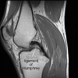
The anterior meniscofemoral ligament of Humphrey produces a localized anterior thickening of the PCL. It usually looks like a small dark lump, such as seen below. It may occasionally be seen as separate from the PCL and mimic an intraarticular body.
MRI KNEE AXIAL
| MRI KNEE AXIAL |
| <embed allowfullscreen="true" flashvars="doc_id=144135162&mem_id=32576475&doc_type=pptx&fullscreen=0&allowdownload=1" height="1428" src="http://widget.wetpaintserv.us/wiki/wikiradiography/widget/docstoc/5c1c670ac603340221941cef3ce80568b8bcae19" type="application/x-shockwave-flash" width="1632" wmode="transparent"/> |
| MRI KNEE CORONAL |
| <embed allowfullscreen="true" flashvars="doc_id=144135166&mem_id=32576475&doc_type=pptx&fullscreen=0&allowdownload=1" height="1428" src="http://widget.wetpaintserv.us/wiki/wikiradiography/widget/docstoc/3891737b6e63bbec0c9dad2f7275fd8636f37a1a" type="application/x-shockwave-flash" width="1632" wmode="transparent"/> |
| MRI KNEE SAGITTAL |
| <embed allowfullscreen="true" flashvars="doc_id=144135168&mem_id=32576475&doc_type=pptx&fullscreen=0&allowdownload=1" height="1428" src="http://widget.wetpaintserv.us/wiki/wikiradiography/widget/docstoc/71462b24d93c8cd9212e15c3107df874daaf972c" type="application/x-shockwave-flash" width="1632" wmode="transparent"/> |
Basic MRI Anatomy Of A Few Parts Of The Knee
Bones
This is a sagittal T1-weighted MR is through the intercondylar notch. The femur and tibia are shown as bright structures (due to the bright marrow fat signal with lots of protons) surrounded by the thin black rim of cortical bone, which has relatively few protons.
anterior
 posterior
 The patella (P) is seen anterior to the femur. The dark structure extending upward from the patella is the quadriceps tendon (q). The dark structure extending caudad from the patella down to the tibial tuberosity is the patellar ligament (p), sometimes called the patellar tendon. If one thinks of the patella as a bone in its own right, then the dark structure (p) coming off of its inferior tip should rightfully be called the patellar ligament, since it connects two bones. However, if you just consider the patella to be a sesamoid bone living in this really long tendon, then calling it the patellar tendon makes some sense.
Suprapatellar Fat Pads
Just above the patella and right behind the quadriceps tendon is the anterior suprapatellar fat pad (a). Just anterior to the femur is the prefemoral fat pad (p). Unlike the anterior suprapatellar fat pad, which is relatively constant in shape and size, this fat pad is quite variable in size, and may appear either fairly flat or quite plump. Extending up between these two fat pads is the suprapatellar bursa, an extension of the knee joint space.
anterior
 posterior
 These two fat pads can usually be seen on a lateral plain film. Usually the suprapatellar bursa is only a potential space, and contains very little fluid. In this case, the two fat pads are separated by less than 5 mm. If a suprapatellar effusion is present, the two fat pads are pushed apart by the effusion. If the distance separating them is 5 mm or greater, we consider this sufficient evidence to diagnose a knee effusion on plain films. We don't use this sign with MR, because we can image the effusion directly.
Cruciate and Anterior Meniscofemoral Ligaments
The anterior and posterior cruciate ligaments are two of the other major ligaments of the knee. Their job is to maintain the anteroposterior relationship of the distal femur and proximal tibia to each other. This job is made a bit trickier by the fact that it needs to be done at all times during flexion or extension.
The anterior cruciate lies lateral to the posterior cruciate. A nice mnemonic for remembering these relative positions is: LAMP, which stands for Lateral Anterior - Medial Posterior.
Posterior Cruciate Ligament
The posterior cruciate is a relatively simple band of tissue and attaches proximally on the internal aspect of the medial femoral condyle and runs distally to where it attaches to the posterior eminence of the tibia. It generally lies in the sagittal plane, and can usually be seen in its entirety on a single sagittal MR slice.

Anterior Cruciate Ligament
The anterior cruciate ligament is usually not a simple linear band. Instead, it is normally fan shaped, as shown below. It attaches proximally at the internal aspect of the lateral femoral condyle, and runs distally to its broad attachment to the anterior tibia and the anterior aspect of the tibial spine.


The anterior cruciate ligament usually lies in a plane 10 - 15° externally rotated to the sagittal plane. Therefore, if one obtains images in this plane, one can image the ACL in over 95% of the time.
Anterior Meniscofemoral Ligament
The anterior meniscofemoral ligament of Humphrey produces a localized anterior thickening of the PCL. It usually looks like a small dark lump, such as seen below. It may occasionally be seen as separate from the PCL and mimic an intraarticular body.
This ligament can be seen a bit better on the enlarged view below.