Lymphangitis Carcinomatosis
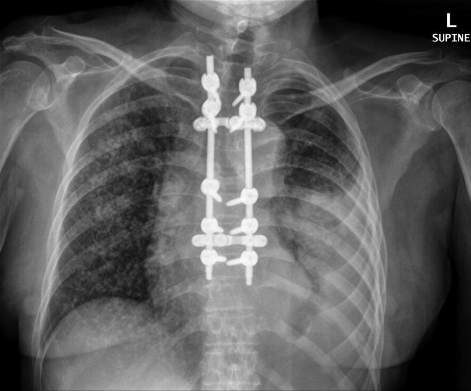
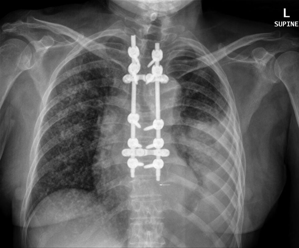
Lymphangitic carcinomatosis (LC) refers to the diffuse infiltration and obstruction of pulmonary parenchymal lymphatic channels by tumour. Various neoplasms can cause lymphangitic carcinomatosis, but 80% are adenocarcinomas. The most common primary sites are the breasts, lungs, colon, and stomach. <a class="external" href="http://emedicine.medscape.com/article/359006-overview" rel="nofollow" target="_blank">(Ali Nawaz Khan, Lymphangitic Carcinomatosis Imaging)</a> The plain film appearance of lymphangitis carcinomatosis (LC) is highly characteristic and therefore readily recognised. It can be useful for radiographers to be aware of the plain film pattern of LC given that there may be other more subtle signs of metastatic disease worthy of supplementary imaging.
Pathology
- Lymphangitic carcinomatosis is believed to initially occur as hematogenous spread of tumor to the lungs
- The tumor then invades the vessels wall and into the lymphatics
- Tumor spreads through the lymphatics which provide little resistance to spread
- Alternatively, there may be central obstruction of mediastinal or hilar lymph nodes leading to direct infiltration
<a class="external" href="http://www.learningradiology.com/archives06/COW+225-Lymphangitic+spread/lymphangiticcorrect.html" rel="nofollow" target="_blank">http://www.learningradiology.com/archives06/COW%20225-Lymphangitic%20spread/lymphangiticcorrect.html</a>
Radiographic Appearance
... back to the wikiradiography home page
... back to the Applied Radiography home page

