Lunate Fractures
Jump to navigation
Jump to search
Introduction
Anatomy
Mechanism of Injury
Presentation
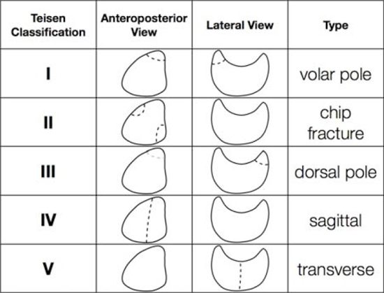
Teisen Classification
Case Study 1
--- under construction----
Introduction
Lunate fractures are uncommon. This page considers all aspects of radiography of these rare fractures.
Anatomy
Mechanism of Injury
- fall onto an outstretched hand
- direct blow
- repetition injury
- secondary to Kienbock's disease
Presentation
"Patients often present with palpation tenderness on the volar wrist. Wrist range of motion is usually painful." <a class="external" href="http://www.orthopaedia.com/display/Main/Lunate+fractures" rel="nofollow" target="_blank">http://www.orthopaedia.com/display/Main/Lunate+fractures</a>
Teisen Classification
Source: <a class="external" href="http://radiology.casereports.net/index.php/rcr/article/viewFile/70/224" rel="nofollow" target="_blank">http://radiology.casereports.net/index.php/rcr/article/viewFile/70/224</a>Teisen classified lunate fractures into 5 groups.
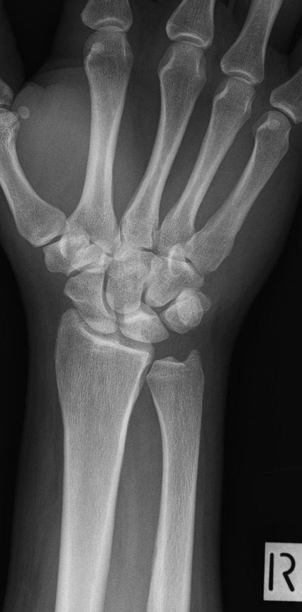
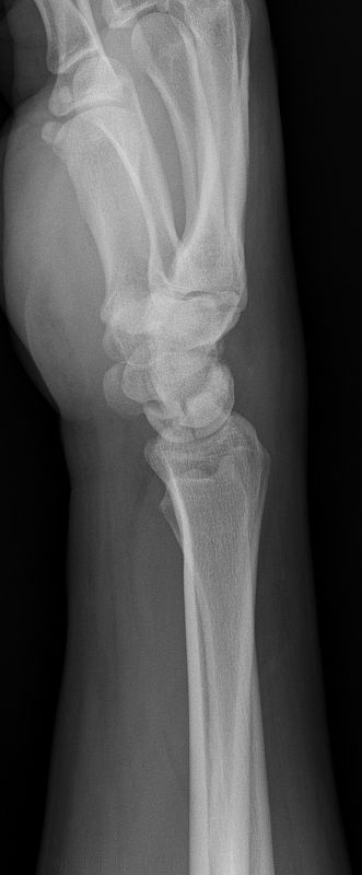
Case Study 1


