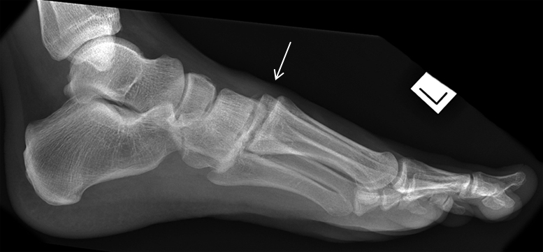
Lisfranc's Fracture
Introduction
The Lisfranc fracture usually appears in lists of commonly missed fractures (up to 20% of Lisfranc injuries are overlooked on initial radiographs). Prognosis can be poor in cases of delayed treatment. A missed diagnosis has serious consequences. An untreated midfoot sprain leads rapidly to osteoarthritis and flattening of the longitudinal arch.
This page considers all aspects of plain film radiography of Lisfranc fractures.
A Little French History
<a class="external" href="http://commons.wikimedia.org/wiki/File:Jacques_Lisfranc.jpg" rel="nofollow" target="_blank">http://commons.wikimedia.org/wiki/File:Jacques_Lisfranc.jpg</a>Named after Jacques Lisfranc(1790-1847), a field surgeon in Napoleon’s army, who described a new technique for an amputation used to treat frostbite of the forefoot in soldiers on the Russian front. <a class="external" href="http://www.learningradiology.com/archives06/COW+217-Lisfranc+fx/lisfranccorrect.htm" rel="nofollow" target="_blank">http://www.learningradiology.com/archives06/COW%20217-Lisfranc%20fx/lisfranccorrect.htm</a>
Relevant Videos
<embed allowfullscreen="true" height="350" src="http://widget.wetpaintserv.us/wiki/wikiradiography/widget/youtubevideo/480d4b5c309fe25dd2471ffd2d8441fe33247a37" type="application/x-shockwave-flash" width="425" wmode="transparent"/> <embed allowfullscreen="true" height="350" src="http://widget.wetpaintserv.us/wiki/wikiradiography/widget/youtubevideo/df8fb52867b23672925d8ca5f9fe4363848e19e0" type="application/x-shockwave-flash" width="425" wmode="transparent"/> <embed allowfullscreen="true" height="350" src="http://widget.wetpaintserv.us/wiki/wikiradiography/widget/youtubevideo/b22441c9d877a7ed520d228139e3a71c70ee3f34" type="application/x-shockwave-flash" width="425" wmode="transparent"/> Could not get this video to embed- click the link to watch the video
<a class="external" href="http://vimeo.com/16013298" rel="nofollow" target="_blank">http://vimeo.com/16013298</a>
Anatomy
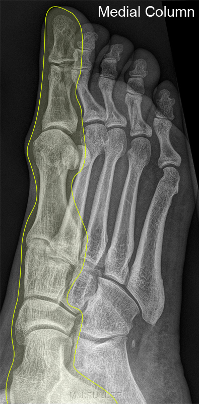
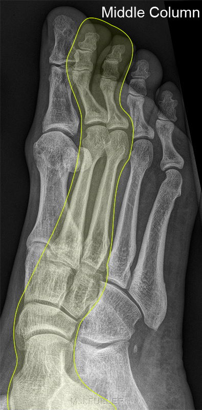
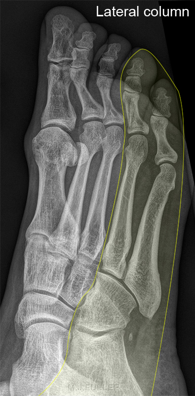
The Columns of the Foot
Medial Column of the foot •1st metatarsal
•Medial cunieform
•Navicular
•TalusLateral Column of the foot •2nd & 3rd metatarsals
•Middle & lateral cunieforms
•Navicular
•TalusLateral Column of the foot Lateral column of foot provides majority of mobility and weight bearing in the foot, Significant alterations in foot biomechanics occur with lateral column joint disruption or loss of length.•4th & 5th metatarsals
•Cuboid
•Calcaneus
Note that the cuboid articulates with the bases of the 4th and 5th metatarsals and the calcaneum.
Nicholas Beckmann, MD and Manickam Kumaravel, MD <a class="external" href="http://www.uth.tmc.edu/radiology/presentations/2008/nutcracker_beckman_2008.pdf" rel="nofollow" target="_blank">Nuts and Bolts of the Nutcracker</a> . http://www.uth.tmc.edu/radiology/presentations/2008/nutcracker_beckman_2008.pdf
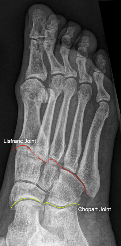
Midfoot Articlations
Midfoot articulations with forefoot and hind foot To lessen ambiguity, some investigators have suggested that the term "Lisfranc joint complex" should be used to refer to tarsometatarsal articulations and that the term "Lisfranc joint" should be applied to medial articulation involving the first and second metatarsals with the medial and middle cuneiforms.•Lisfranc joint (forefoot articulation, red line)
•Chopart joint (hind foot articulation, yellow line)<a class="external" href="http://www.radsource.us/clinic/0908" rel="nofollow" target="_blank">quoted from </a>
<a class="external" href="http://www.radsource.us/clinic/0908" rel="nofollow" target="_blank">MRI Web Clinic - August 2009</a>
<a class="external" href="http://www.radsource.us/clinic/0908" rel="nofollow" target="_blank">Lisfranc Ligament Tear</a>
<a class="external" href="http://www.radsource.us/clinic/0908" rel="nofollow" target="_blank">by Gary A. Howell, M.D.</a>
Mechanism of Injury
<a class="external" href="http://www.radsource.us/clinic/0908" rel="nofollow" target="_blank"> </a>
<a class="external" href="http://www.radsource.us/clinic/0908" rel="nofollow" target="_blank">http://www.radsource.us/clinic/0908
</a><a class="external" href="http://www.radsource.us/clinic/0908" rel="nofollow" target="_blank">
</a>
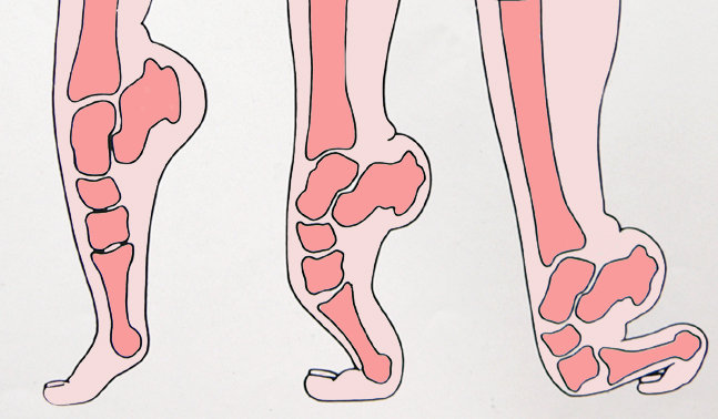
adapted from Source: Harris, JH Jr, Harris WH, Novelline, RA, The Radiology of Emergency Medicine Third Ed.p986 Willams and Wilkins, 1981Lisfranc joint fracture dislocations and sprains can be caused by high-energy forces in motor vehicle crashes, industrial accidents and falls from high places.Occasionally, these injuries result from a less stressful mechanism, such as a twisting fall. Since Lisfranc joint fracture dislocations and sprains carry a high risk of chronic secondary disability, physicians should maintain a high index of suspicion for these injuries in patients with foot injuries characterized by marked swelling, tarsometatarsal joint tenderness and the inability to bear weight.
HIGH-VELOCITY INJURY
This type of injury to the Lisfranc joint typically occurs with major, high-velocity trauma such as motor vehicle accidents or falls from a height. The force on the Lisfranc joint is abduction and/or plantar flexion. The joint is grossly dislocated, and fractures of the metatarsals or cuneiforms are commonly seen.
Fracture dislocations are classified as either homolateral with all five metatarsals displaced in either the medial or lateral direction, or divergent, where a separation of the first and second metatarsals occurs.
LOW-VELOCITY INJURY: MIDFOOT SPRAIN
Low-velocity injuries can also result in mechanically significant disruption of the Lisfranc joint. Two injury mechanisms have been described. The first mechanism is plantar flexion and axial load, sometimes with a component of inversion. It occurs when the patient's foot is plantar flexed and suffers an axial load injury. This injury may occur during running and jumping sports or when a patient trips going downstairs or stepping off a curb. It also may occur when a downward force is applied to the heel when the patient's knee is on the ground and the ankle is dorsiflexed.
The second mechanism is forced forefoot abduction. This injury may also occur when an athlete wearing cleats plants the foot and turns quickly. In both mechanisms of injury, stress is applied to the Lisfranc ligament complex, and ligament failure, referred to as "midfoot sprain", occurs. Midfoot sprain is uncommon in non-athletes, but in athletes it is the second most common athletic foot injury, occurring in 4% of football players per year.<a class="external" href="http://www.radsource.us/clinic/0908" rel="nofollow" target="_blank">quoted from </a>
<a class="external" href="http://www.radsource.us/clinic/0908" rel="nofollow" target="_blank">MRI Web Clinic - August 2009</a>
<a class="external" href="http://www.radsource.us/clinic/0908" rel="nofollow" target="_blank">Lisfranc Ligament Tear</a>
<a class="external" href="http://www.radsource.us/clinic/0908" rel="nofollow" target="_blank">by Gary A. Howell, M.D.</a>
The Lisfranc Fracture
Used today to describe fractures and dislocations that occur at the junction between the tarsal bones of the midfoot and the metatarsals of the forefoot
Radiography of Lisfranc Injuries
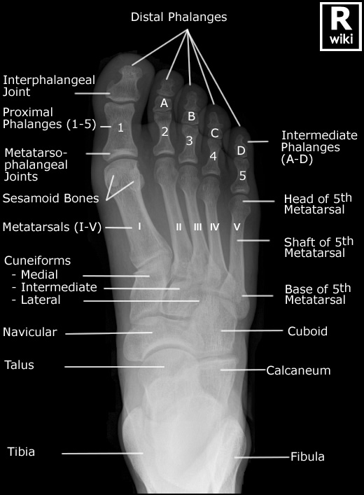
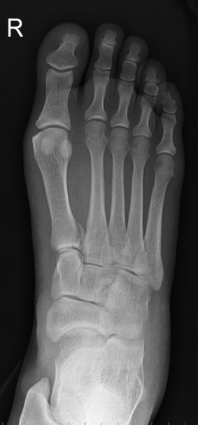
AP/DP Foot
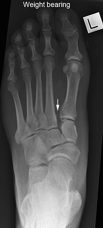
Weightbearing AP/DP Foot
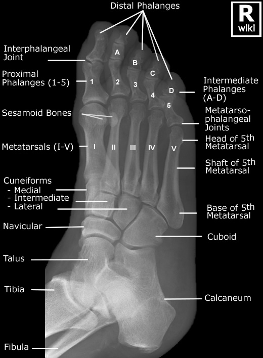
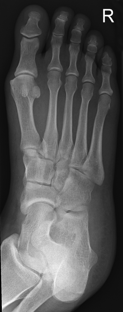
Oblique Foot
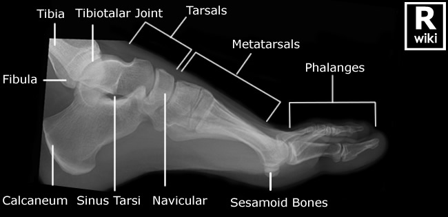
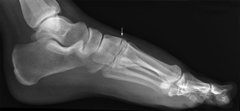
Lateral

