Left Lower Lobe Consolidation
Introduction
Left Lower lobe (LLL) is a relatively common site for consolidation and can be a tricky diagnosis if the image is underpenetrated and/or if the consolidation is not very dense and/or if a lateral view is not included in the series.
The Meaning of the Term Consolidation
One of the unfortunate aspects of the term consolidation is that its meaning can be different depending on who is using the term. When a clinician uses the term consolidation he/she is usually referring to a consolidation associated with acute pneumonia. Thus, the term consolidation and pneumonia have very similar meanings and are almost used interchangeably.
Strictly speaking, the term consolidation does not imply any particular aetiology or pathology. Acute pneumonia is the commonest cause but not the only cause of consolidation. ( other causes include chronic pneumonia, pulmonary oedema and neoplasm). Thus when a radiologist has reported a chest X-ray examination and notes the presence of consolidation he/she is simply stating that some of the long airspace has been replaced by a fluid.
The Left Lower Lobe Anatomy
adapted from <a class="external" href="http://books.google.com.au/books?id=Bif0zpmEWtAC" rel="nofollow" target="_blank">By Fred W. Wright Radiology of the Chest and Related Conditions: Together with an Extensive Illustrative Collection of Radiographs CRC Press, 2002</a>The left lower lobe is similar in structure to the right lower lobe except that it has two segments combined- because the anterior and medial basal segments share a common bronchial supply, these two segments are characteristically combined, forming an anterior medial basal segment. More information on lung anatomy here
adapted from <a class="external" href="http://books.google.com.au/books?id=Bif0zpmEWtAC" rel="nofollow" target="_blank">By Fred W. Wright Radiology of the Chest and Related Conditions: Together with an Extensive Illustrative Collection of Radiographs CRC Press, 2002</a>


Notes on Consolidation
- Consolidation refers to fluid in the airspaces of the lung
- Consolidation may be complete or incomplete
- The distribution of the consolidation can vary widely. A consolidation could be described as “patchy”, “homogenous”, or generalised”.
- A consolidation may be described as focal or by the lobe or segment of lobe affected
Plain Film Appearances of Lung Consolidation
Radiological appearances common to all lobes are:
1.Abnormal lung opacity2.Increase in the size and number of lung markings3.Loss of clarity of the diaphragm on the AP and/or lateral views4.Loss of clarity of the heart border on the AP and/or lateral views5.Air bronchogram lines6.Loss of the normal darkening inferiorly of the thoracic vertebral bodies on the lateral view7.Opacification of the lung behind the heart shadow or below the diaphragms
Look Behind the Heart Shadow
One of the more subtle appearances of consolidation can be seen when the left heart shadow appears abnormally dense. Benjamin Felson (<a class="external" href="http://www.amazon.com/Chest-Roentgenology-Benjamin-Felson/dp/0721635911/ref=sr_1_2?ie=UTF8&s=books&qid=1252240078&sr=1-2" rel="nofollow" target="_blank">Chest Roentgenology, W.B. Saunders, 1973, p23</a>) notes that "... the cardiac thickness is approximately the same on both sides of the spine; thus the heart normally casts a uniform roentgen shadow over its entire area".
Obliteration of the Descending Aorta"The descending aorta indents the superior and posterior basal segments of the LLL, and its lateral margin is therefore obliterated by lesions in these segments". Benjamin Felson (<a class="external" href="http://www.amazon.com/Chest-Roentgenology-Benjamin-Felson/dp/0721635911/ref=sr_1_2?ie=UTF8&s=books&qid=1252240078&sr=1-2" rel="nofollow" target="_blank">Chest Roentgenology, W.B. Saunders, 1973, p36</a>)
•Appears as an area of increased opacity within the LLL•Some loss of the hemi-diaphragm is commonly seen•May be increased density behind left heart shadow
Note- if you are having trouble understanding why a lung density that is clearly extending into the upper part of the patient's chest is contained within the lower lobe, think about the actual coverage of the left lower lobe- it extends quite high in the chest. (see the lateral chest graphic below)•Appears as an area of increased opacity within the LLL•Some loss of the hemi-diaphragm medially is seen•increased density behind left heart shadow
•Increased opacity within the LLL•Commonly seen with loss of the Left hemi-diaphragm•May be sharply delineated by oblique fissure
•Increased opacity within the LLL•Loss of the normal darkening of the thoracic spine inferiorly
- some loss of the left hemi-diaphragm posteriorly


Single View Chest Radiography
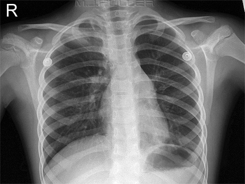
This is a PA/AP chest image on a paediatric patient. It may be tempting in a difficult patient to finish the examination at this point. The lung fields are well demonstrated and you can even see behind the heart and under both hemi-diaphragms. This is a lateral chest image on the same patient. Note the left lower lobe consolidation. In retrospect, there is increased opacity seen behind the heart on the AP view, but this could easily be missed.
Any single view radiography is hazardous, and the chest is no exception.
There is further discussion on the value of a lateral chest image here.


Subtle LLL Consolidation
This 18 year old male presented to the Emergency Department with headache, fever and viral signs. There is increased opacity of the left lung behind the heart shadow. This is easier to appreciate when compared with the normal CXR taken 2 years earlier (right) This CXR from two years earlier demonstrtaes a normal LLL. The lateral view similarly demonstrates a very subtle sign of consolidation. The thoracic vertebral bodies should show an evenly graduated darkening from the top to bottom. This image demonstrates a few lightly lightened thoracic vertebral bodies behind the heart. Compare with the lateral chest image taken a few yars earlier. Normal lateral chest X-ray taken a few years earlier.


Case 1
There is a left sided pleural effusion (grey arrow). There is greater density below the left hemidiaphragm than the right (black and white arrows respectively). This is a very subtle sign of chest pathology on a PA chest image The right hemidiaphragm is visualised and the left hemidiaphragm is largely obscured (silhouette sign). There is LLL density which is likely to be a combination of effusion, collapse and consolidation.


Case 2
There is abnormal density within the left lower lobe which is sharply marginated medially (white arrow). Note that the heart appears darker to the right of the spine compared to the heart visible to the left of the spine.
There is loss of the left hemidiaphragm known as silhouette sign (black arrow)
There are also airbronchogram lines (not marked)
There is obliteration of the descending aorta.The right hemidiaphragm is clearly visualised.
The left hemidiaphragm is not seen (silhouette sign)
The air/fluid level is within the stomach (white arrow)
There is abnormal density demonstrated posteriorly. This is causing the lower thoracic vertebral bodies to demonstrate a decreased density. This is an important sign of disease on the lateral image. The vertebral bodies should appear to darken evenly as you look from the upper vertebral bodies down to the lower vertebral bodies. This case demonstrates the opposite effect- the vertebral bodies appear lighter as you look from top to bottom.


Case 3
This is a 60 year old patient who presented to the Emergency Department with a worsening productive cough.
There is increased abnormal lung density behind the heart shadow. The heart is a homogenous organ- it should not normally appear to have a different density on the left side of the spine compared to the right. This is a common sign of left lower lobe lung pathology. There is no convincing loss of clarity of the left hemidiaphragm. The anterior part of the hemidiaphram is the part that you see profiled on a PA/AP chest image. The fact that there is no diaphragmatic silhouette sign on the PA image suggests that the pathology is positioned more posteriorly in the RLL.There is increased density behind the heart shadow (arrowed). This causes loss of the normal darkening of the lower thoracic vertebral bodies. This is a common and reliable sign of lower lobe lung pathology. Note that the pathology is not involving the anterior segments of the left lower lobe. This explains why there is no loss of the visible profiled anterior hemidiaphragm on the PA image.


...back to the Applied Radiography home page