Lateral Chest Case 4
This is the answer page to Case 4 from the page titled What is the Value of the Lateral Chest Projection?
Other Relevant Wikiradiography Pages
- What is the Value of the Lateral Chest Projection?
- Patterns of Consolidation
- Patterns of Consolidation- LUL
- Lung Anatomy
- Paediatric Chest Radiography Positioning Aids
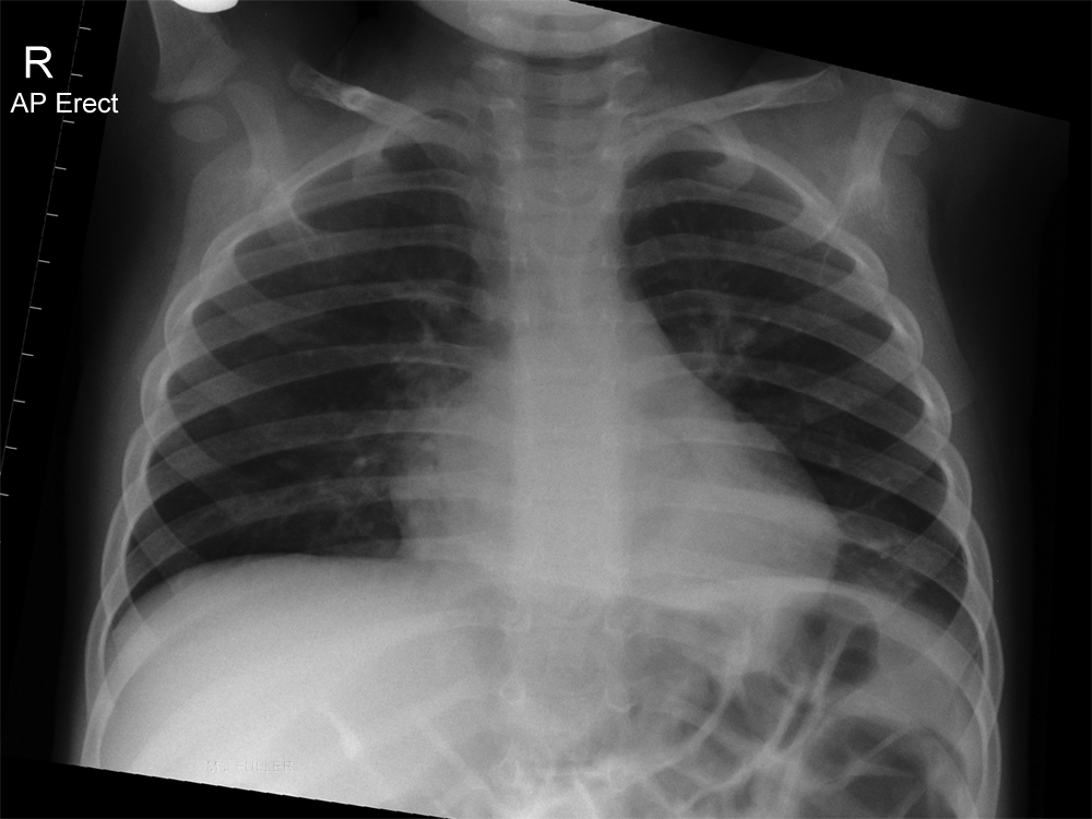
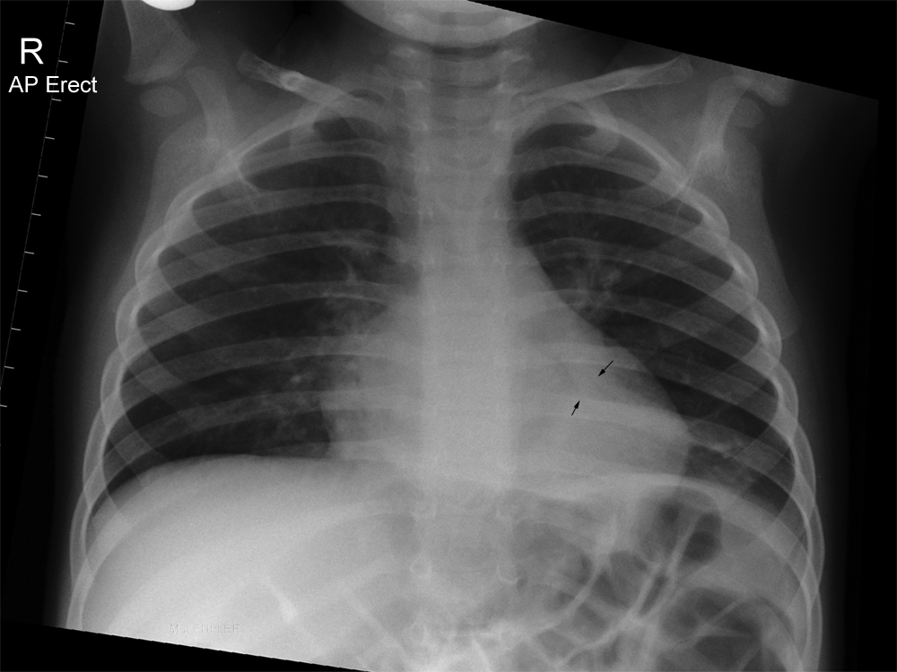
AP sitting Projection
Unlabelled
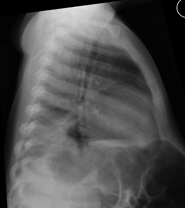
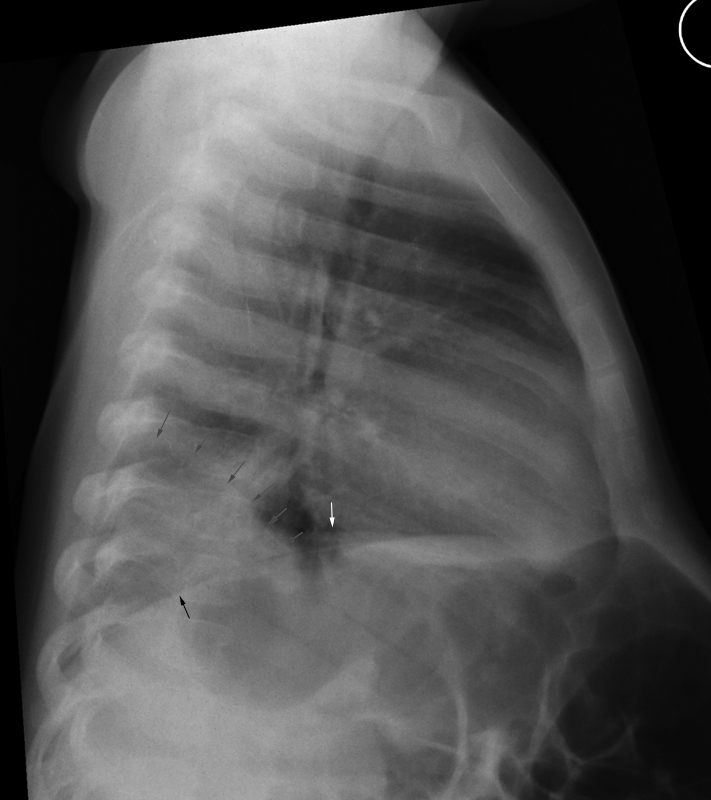
Lateral
The right hemidiaphragm is demonstrated (black arrow)
The left hemidiaphragm is demonstrated partially (white arrow) and becomes obscured posteriorly.
There is abnormal opacity overlying the lower thoracic spine region (grey arrows)
Discussion
This child has a left lower lobe consolidation which is likely to represent an acute pneumonia. The significant clue on the AP projection image is the presence of airbronchogram lines behind the heart. Whilst this condition can be diagnosed from the AP sitting projection by any person with skills in image interpretation, a less skilled observer could easily have missed this finding if a lateral projection image was not available. This is the sort of patient that turns up to the Emergency Department three days later in a considerably worse state than they presented the first time.
The pneumonitis is much easier to diagnose on the lateral projection image. There is abnormal posterior basal opacity with a silhouette sign involving the left hemidiaphragm. Another significant clue is that the thoracic spine should not appear lighter as you look from top to bottom- if anything, it should appear slightly darker as you look down the spine (until you reach diaphragm)
If this was your patient, would they have gone home with an undiagnosed left lower lobe pneumonia?... back to the wikiRadiography home page
... back to the Applied Radiography home page
... back to the What is the Value of the Lateral Chest Projection? page


