Lateral Chest Case 3
Jump to navigation
Jump to search
Preamble
Other Relevant Wikiradiography Pages
... back to the Applied Radiography home page
... back to the What is the Value of the Lateral Chest Projection? page
This is the answer page to Case 3 from the page titled What is the Value of the Lateral Chest Projection?
Other Relevant Wikiradiography Pages
- What is the Value of the Lateral Chest Projection?
- Patterns of Consolidation
- Pneumoperitoneum
- Pneumoperitoneum- Radiographic Techniques
PA Chest
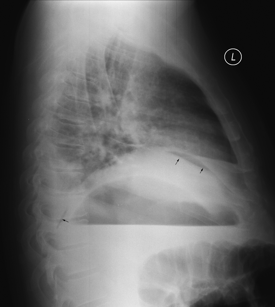
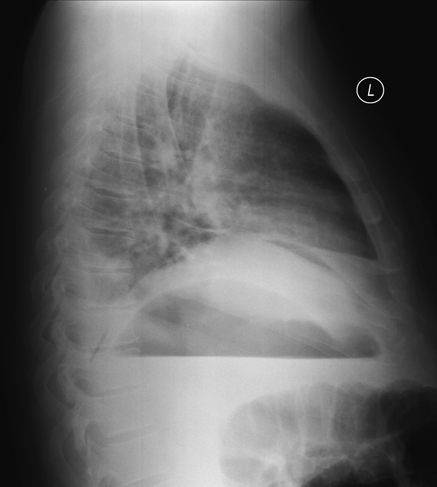
The lateral projection image demonstrates air interposed between the liver and diaphragm
 |
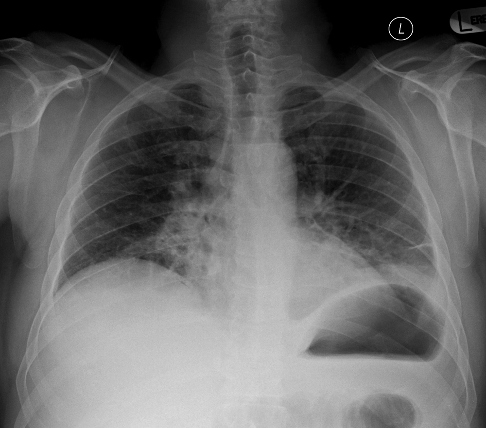
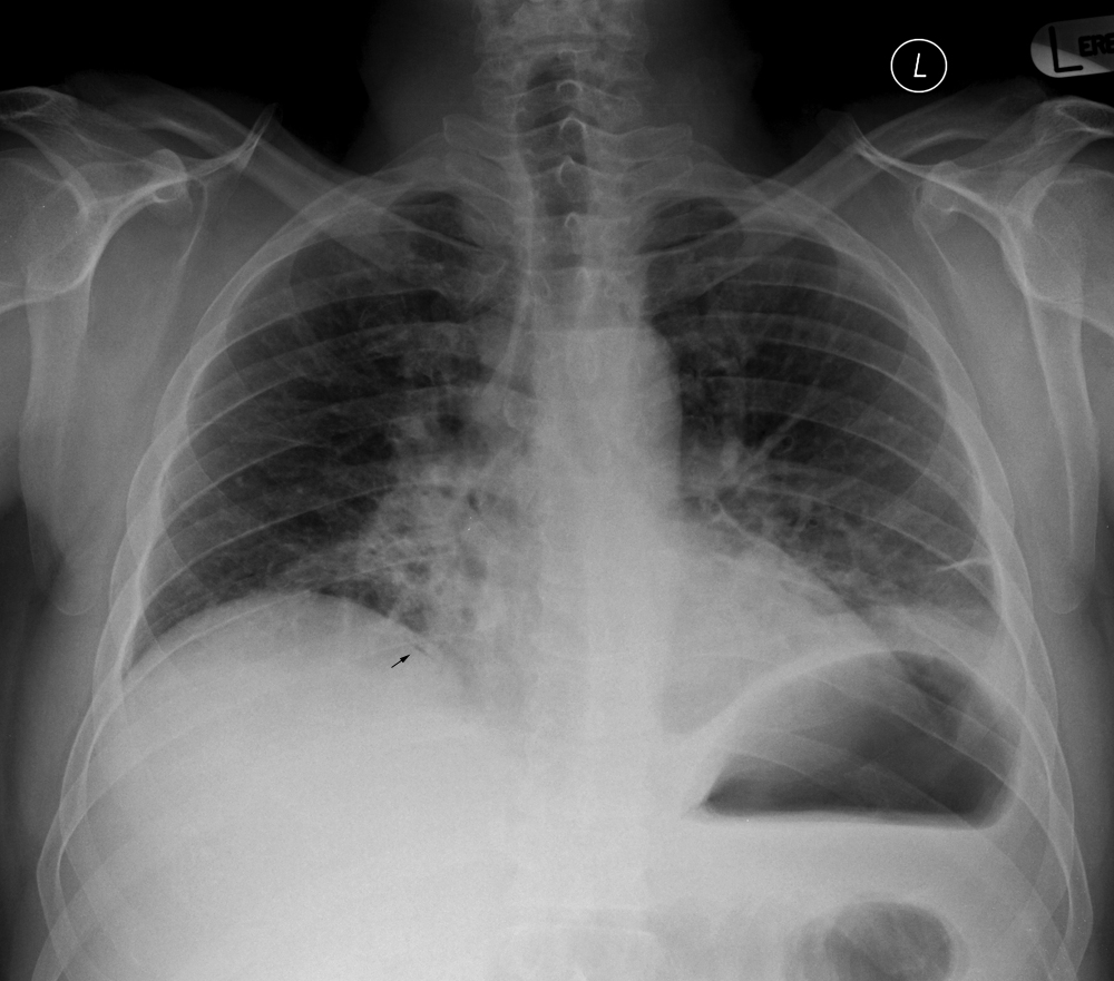
| There is a large air/fluid level in the stomach The lateral projection image demonstrates air interposed between the liver and diaphragm (arrowed) This appearance indicates pneumoperitoneum from perforated hollow abdominal visus. Contrary to popular belief, research findings and the author's experience suggest that the lateral chest centred on the diaphragm is the most sensitive projection for detection of pneumoperitoneum. see Pneumoperitoneum and Pneumoperitoneum- Radiographic Techniques |
... back to the Applied Radiography home page
... back to the What is the Value of the Lateral Chest Projection? page


