Introduction Bowel obstructions are common abdominal pathologies. This page considers normal appearances of the large bowel and patterns associated with large bowel obstruction. A few conditions that mimic large bowel obstruction (LBO) are also covered. There are a number of pages on this wiki that cover various aspects of abdominal plain film imaging listed here. It is worth reading the definition of terms page before this page.
Definition of Terms Obstruction
Bowel obstruction refers to a mechanical obstruction to the lumen of the bowel causing stasis of the bowel contents above a focal lesion
•Complete or partial
•Cause- intrinsic, extrinsic, intra-luminal
Closed Loop Obstruction
Obstruction of the bowel at two separate points produces a closed loop. A gas-filled closed loop may double on itself and assume a ‘U’ shape resembling a coffee bean- the
Coffee bean sign.
Normal Radiological Appearance
| - Large bowel is characterized on plain film by its haustrations and sacculations. These are most prominent in the ascending and transverse colon but can be seen in the left colon.
- With moderate distention of the large bowel, the plicae appear to extend entirely across the lumen but this appearance may disappear with further distension.
- The plicae of the large bowel are more widely spaced than the valvulae of the small bowel.
- Large bowel will normally contain solid material- small bowel almost always contains liquid and gas only. Solid faeces always conclusively identifies the colon.
- Large bowel tends to be peripherally located. (beware the sigmoid)
- Normally, the large bowel contains at least a small amount of gas and, in the supine position, the least dependant (most anterior) segment is the transverse colon followed by the sigmoid.
- The identification of the transverse colon will usually assist in identifying the position of the stomach which lies above it (the gastrocolic ligament is variable in length)
- Fluid levels in the abdomen are seen in normal patients commonly in the stomach, often in the small bowel and never in the colon distal to the hepatic flexure (Stephen Baker, The Abdominal Plain Film)
- Occasionally the features of large or small bowel are not distinct and a loop of bowel is demonstrated that has features of both.
|
|
Causes of LBO
- Mechanical obstruction of the large intestine is most often due to primary carcinoma of the colon (60%).
- Post-surgical adhesive bands rarely block the large bowel lumen.
The String of Pearls Sign 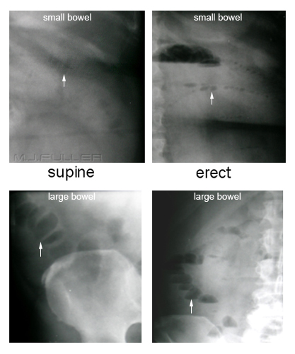 | The string of pearls sign is a distinct appearance associated with small bowel obstruction. There is however a similar appearance that can occur in the large bowel. The large bowel "pearls" are bigger than those of the small bowel and tend to have flat bases |
Radiological Appearances of LBO 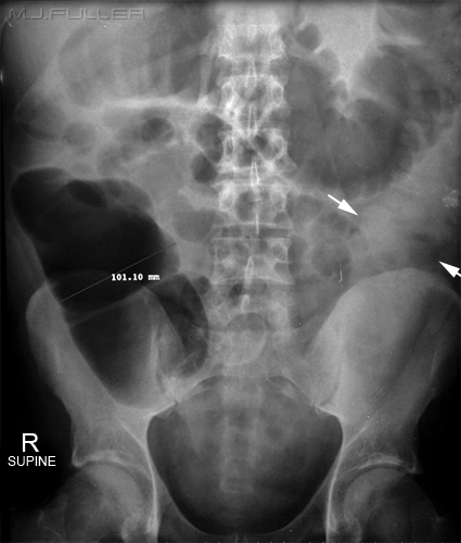 | - Approximately 25% of all intestinal obstructions occur in the large bowel
- Gas and faeces tend to accumulate proximal to the point of obstruction.
- In a typical configuration of mechanical obstruction, all colonic segments proximal to the point of luminal narrowing are dilated.
- In most cases of large bowel obstruction, the bowel will contain variable amounts of solid, liquid and gaseous constituents.
- Fluid levels in the large bowel tend to be less in number but longer than those seen in the small bowel.
- Completely fluid-filled large bowel may go undetected on plain films (white arrows).
- In long-standing LBO, muscular exhaustion can ensue, resulting in the effacement of intra-luminal septa and haustra.
- The colon is dilated when it exceeds 6cm in diameter, and the caecum is dilated when it exceeds 9cm in diameter. (3,6,9 rule)
- When the caecal diameter exceed 10cm, the probability of perforation is high.
- The caecum always dilates to the largest extent no matter where the LBO is sited (Laplace’s law ).
|
Laplace's Law Case 1
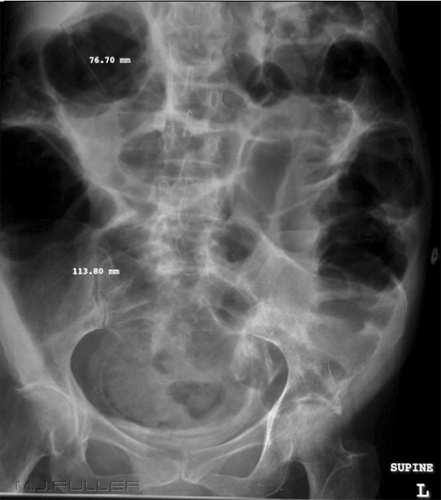 | Laplace’s Law
If there is free flow of gas and fluid along a segment of bowel, intra-luminal pressure must be constant throughout. Laplace’s law states that the pressure needed to distend a hollow viscus varies inversely with its radius. Thus, the caecum, usually the widest part of the colon, expands to the greatest extent as the large bowel dilates. Case 1 This case shows a distended transverse colon (77mm) and a distended caecum (113mm). Laplace's Law would suggest that this is a genuine case of colonic obstruction. This was proven with subsequent enema which demonstrated an obstruction of the distal sigmoid. 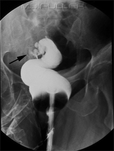 |
Case 2
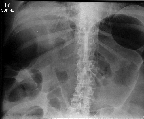 | Case 2
This patient demonstrated a grossly dilated transverse colon. Note that the caecum appears to be of normal calibre. The enema image shows a smooth transition from transverse colon to splenic flexure suggesting that the cause of the massively dilated transverse colon is not obstruction. This appearance is typical of colonic pseudo-obstruction.
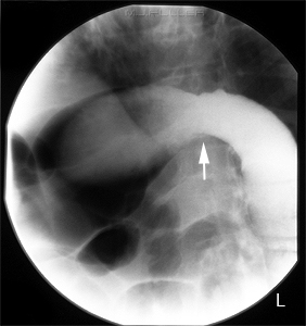 |
|
|
Faecal Impaction and LBO
| Faecal impaction is a common occurrence in : - the elderly (most common cause of colonic obstruction)
- neurologically impaired
- bed-ridden
- younger patients who are chronic abusers of narcotic medications.
The serious consequences of intractable constipation in the aged or infirm is sometimes underestimated (blood flow/abrasive effects). |
|
The 3,6,9 Rule  | - The colon is dilated when it exceeds 6cm in diameter, and the caecum is dilated when it exceeds 9cm in diameter. (3,6,9 rule)
- The caecum always dilates to the largest extent no matter where the LBO is sited. (Laplace’s law )
- When the caecal diameter exceed 10cm, the probability of perforation is high
|
The Air in the Rectum Conspiracy 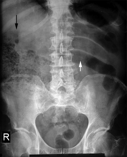 | - Obstruction of the bowel does not mean immediate disappearance of distal gas
- A partial obstruction will allow passage of air distally
- In cases of complete obstruction, the fermentation of residual faecal matter in the large bowel can produce gas distal to the obstruction for some time after the bowel becomes completely obstructed
This patient has a tight stricture at the level of the hepatic flexure. Despite this stricture, there is clearly gas in the rectum. |
|
|
Establishing the Level of Obstruction
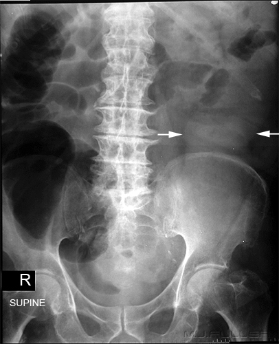 | - The exact point of a large bowel obstruction is difficult to detect on abdominal plain films. In most cases, the location of the terminus of the colonic gas shadow does not necessarily correspond with the site of the bowel occlusion.
- A typical pattern is continuous dilation of the caecum, right colon, and transverse colon up to the splenic flexure caused by an obstructing tumour in the sigmoid colon. The intervening descending colon lacks gas, as it is distended by fluid and solid faeces only.
This is a supine abdominal image on a patient who presented to the Emergency Department with acute abdominal pain. There is a dilated air-filled caecum and ascending colon. The transverse colon is not visualised. The level of obstruction might be assumed to exist in the transverse colon, however, close examination shows the descending colon is also dilated (white arrows) but is less obvious because it is filled with fluid rather than air. 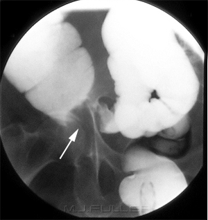 The patient proceeded to have a gastrografin enema which demonstrated an applecore lesion in the sigmoid colon (white arrow). |
|
|
LBO Posing as an SBO  | At a cursory glance this patient appears to have a SBO. On closer examination, the prominent air-filled loops of small bowel in the LUQ (white arrow) have the features of ileum rather than jejunum. Also, the caecum appears unusually large and there appears to be a sudden change in calibre in the large bowel at the level of the hepatic flexure(black arrow). This was reported as a SBO. An alternative explanation is that the large bowel is obstructed at the level of the black arrow. This would account for the dilation of the caecum. An LBO in patients with incompetent ileocaecal valves can mimic an SBO. The ileal loops may have been displaced by the enlarged caecum or they may be effaced jejunal loops..
Note that the patient has the reliably unreliable sign of gas in the rectum!
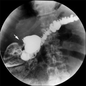
This is a barium enema on the same patient. Note the tight apple-core lesion at the level of the hepatic flexure (white arrow)
Approximately a quarter of patients have an incompetent ileocaecal valve. |
Generalised Adynamic Ileus 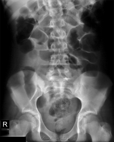 | The bowel could reasonably be said to be a very sensitive organ. It has a propensity to stop functioning with little provocation. Amongst the possible causes are infection(anywhere), abdominal inflammation , chemical/pharmacological causes and trauma.
Abdominal surgery commonly results in generalised adynamic ileus in which the bowel is temporarily non-functioning. This typically manifests on around day 4 post-op. In response, the patients are often referred for abdominal plain film imaging to rule out obstruction.
The appearance of generalised adynamic ileus is quite characteristic. The large and small bowel are extensively airfilled but not dilated. I have heard this described as the large and small bowel "looking the same". |
Caecal and Sigmoid Volvulus
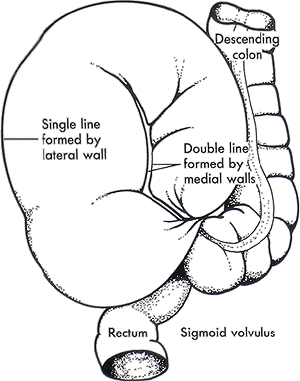
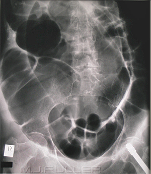
| Caecal Volvulus | Sigmoid Volvulus |
|  source unknown source unknown |
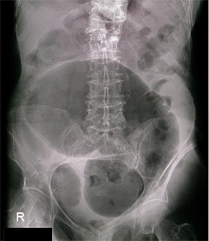 |  |
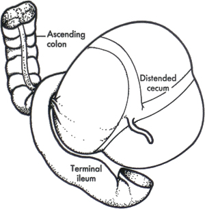
Caecal Volvulus
- caecum is characteristically relocated to the mid-abdomen or left upper quadrant
- characteristically, the walls are smooth and the haustra are preserved
- Persistent dilated distal colon is rarely seen
|
Sigmoid Volvulus
- extends into the right upper abdomen to T10 or higher
- The colon proximal to the twist distends
- The rectum usually empties
- Gas distended sigmoid usually shows Coffee bean sign in common with other closed loop obstructions
- At the point of the twist a barium enema demonstrates a characteristic beak-like termination
|
Colonic Pseudo-obstruction

 -Grossly dilated transverse colon on supine plain film -Grossly dilated transverse colon on supine plain film
-normal caecum
-smooth transition to normal calibre splenic flexure on enema
-probably colonic pseudo-obstruction | Causes - Idiopathic/unknown (acute cases sometimes referred to as Ogilvie’s syndrome)
- A severe form of pseudo-obstruction is sometimes found in mentally disabled and psychotic individuals
Features - No other physical abnormalities
- Rapidly progressive dilation of the colon
- Uncontrolled by rectal tube or colonic decompression
- Untreated- may lead to perforation and death
Pattern Recognition - In most cases, the plain film appearance is sufficiently characteristic to differentiate it from large bowel obstruction
- Haustra are smooth, regularly spaced, and the septa are smooth, thin and sharply marginated
- Lumen is gas filled and the wall contour is clearly demarcated (as distinct from obstruction in which shaggy interfaces are often seen)
- Can be confirmed by gastrografin enema but this is usually unnecessary
|
|
|
|
|
Post Washout and Post Endoscopy 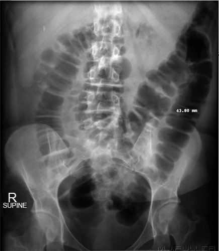 | The large bowel in this image looks unusual and it is not immediately obvious why. The unusual appearance could be attributed to two features. The first is that the colon is extensively air-filled and the small bowel is not. The second feature is that the large bowel contains no faeces. This is an appearance that you will occasionally see in patients who have had bowel washouts. Note that at 44mm diameter, the colon is not enlarged. |
Ischaemic Bowel 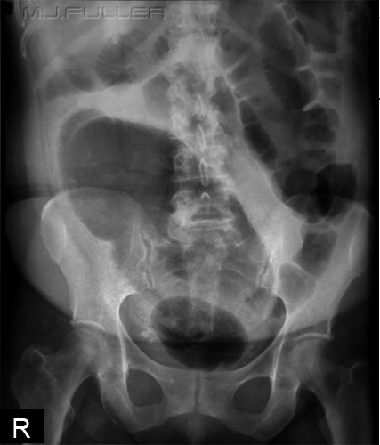 | This patient has a very dilated rectum caused by ischaemia. Where there is an isolated unexplained segment of grossly dilated colon ischaemia should be considered.
? early signs of mural gas |
Case 1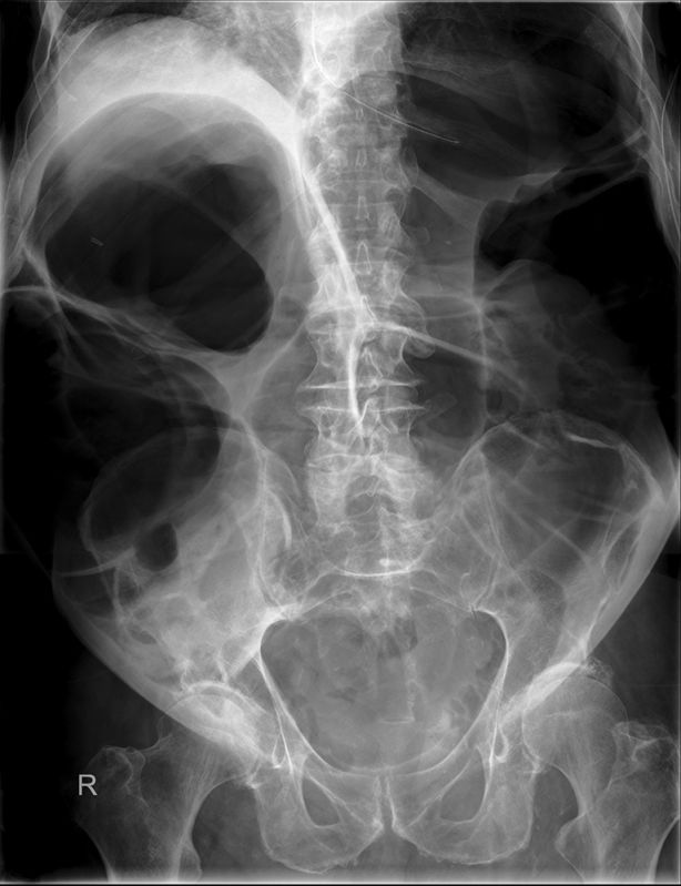 | This 88 year old man presented to the Emergency Department with abdominal pain and distention. The patient was examined and refered for an acute abdominal plain film series. A NGT had been inserted at the time of imaging.
All of the large bowel is air-filled and distended. The appearance is more typical of large bowel obstruction in the presence of a competent ileo-caecal valve than it is of pseudo-obstruction. |
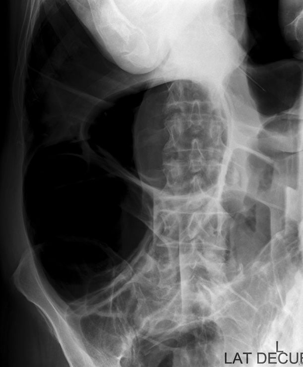 | The left lateral decubitus image demonstrates a distended gas-filled caecum and multiple air-fluid levels. |
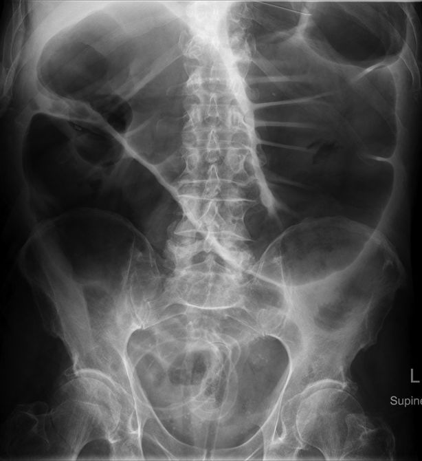 | The patient was referred for colonoscopy and a flatus tube was inserted during the procedure. Follow up abdominal plain film imaging demonstrated some reduced bowel calibre following the flatus tube insertion. |
Summary Colonic obstruction has significant associated morbidity and mortality if untreated. Timely diagnosis on plain abdominal film will potentially improve patient prognosis. The abdominal plain film may not present a radiographic challenge for a seasoned radiographer but can present an interpretation challenge for radiographer and radiologist alike. Radiographer skills in image interpretation will afford additional meaning and professional satisfaction.
......back to the applied radiography home page


