Knee Dislocations and Subluxations
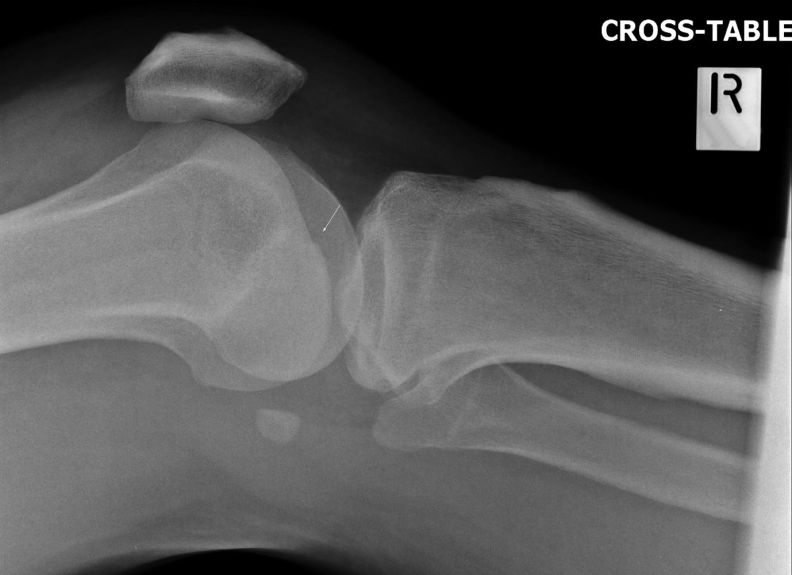
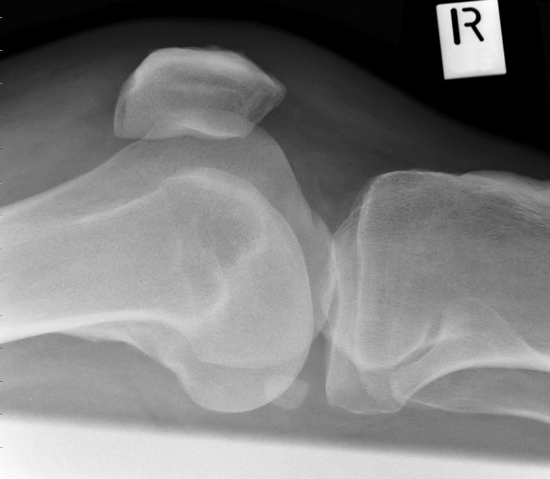
True complete dislocations of the knee joint are uncommon but not rare. This page considers radiography of knee dislocations as well as dislocations and subluxations of the patella and fibula head.
Complete Knee Dislocation Associated Soft Tissue Injuries
Complete Knee Dislocation ClassificationsKnee dislocations are associated with significant soft tissue injury. The following structures are commonly involved
- Anterior cruciate ligament
- posterior cruciate ligament
- joint capsule
- lateral collateral ligament
- medial collateral ligament
- popliteal artery
- biceps femoris tendon
- iliotibial band
- popliteus tendon
- miniscii
<a class="external" href="http://www.wheelessonline.com/ortho/traumatic_dislocations_of_the_knee" rel="nofollow" target="_blank">Wheeless</a> describes the following classifications of knee dislocations
- classification: 5 types: described with tibia in relation to femur;- anterior (31%)
- posterior (25%)
- lateral (13%)
- medial ( 3%)
- rotary ( 4% - usually posterolateral)
The Role of MRI
MRI scanning of the knee following relocation will provide information to assist the surgeon to reconstruct the soft tissue structures of the knee.
Treatment
Surgical repair is usually delayed for one week. There is some debate regarding surgical repair vs conservative management with immobilisation.
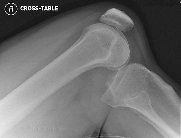
Anterior Dislocation of the Knee
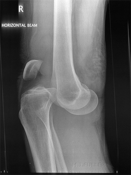
Posterior Knee Dislocation
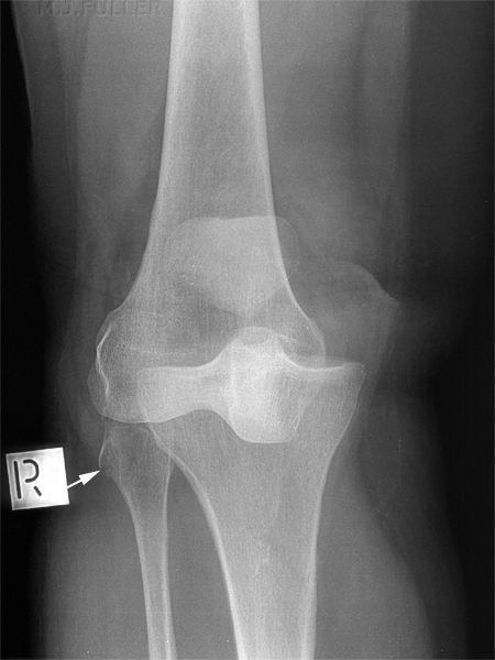
The fracture of the fibula head may appear trivial and incidental in the context of such a serious joint disruption. This is not the case. This patient had his fibula head fracture reduced and screwed back to the fibula to improve his outcome. Failure to re-attach the fibula fracture fragment (and associated soft tissue structures) may contribute to a varus deformity of the knee. Importantly, if the fracture has been reduced in the field, or reduced spontaneously, the fibula fracture known as arcuate sign is a significant finding.
Avulsion fracture of the fibular head, referred to as the arcuate sign, indicates an injury to at least one of the posterolateral corner structures of the knee ...These ligamentous and tendinous structures have been variably termed the arcuate complex. Inserting from medial to lateral on the fibular head, they include the popliteofibular ligament (also known as the fibular insertion of the popliteus muscle), the arcuate ligament, and the conjoined tendon formed by the biceps femoris muscle tendon and fibular collateral ligament. The variably present fabellofibular ligament, posterolateral joint capsule, lateral gastrocnemius muscle, and popliteus muscle are also considered part of the complex .... we propose that the arcuate sign be considered not only as a marker of internal knee derangement, but also of potential knee dislocation.
Jason T. Crimmins, M.D., and Robert D. Wissman, M.D.
The Arcuate Sign: A Marker of Potential Knee Dislocation? A Report of Two CasesRadiology Case Reports,Vol 3, No 2 (2008). <a class="external" href="http://radiology.casereports.net/index.php/rcr/issue/view/13" rel="nofollow" target="_blank">Vol 3, No 2 (2008)</a>
Case 1
Discussion
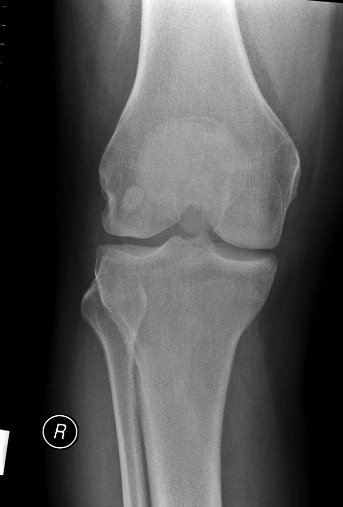
The trauma radiographer should be aware of the arcuate sign and its significance. Subluxation of the proximal fibula is uncommon and may be missed on plain film. Comparison views of the contra-lateral knee may be justified to confirm the diagnosis.
... back to the Applied Radiography home page
