Knee: non-trauma soft tissue signs and artifacts
Jump to navigation
Jump to search
Prepatellar Bursitis
Varicose Veins
This is largely an incidental finding. Varicose veins can produce a distracting and confusing appearance to the uninitiated.
These varicose veins appear to hang down like grapes on a vine.

The Blanket Artifact
In an act of desperation radiographers will roll up a blanket and use it as a support for the lateral horizontal ray knee projection. The result is commonly less than ideal as shown below
There is moderately severe extra-capsular soft tissue swelling (top white arrow). Hoffa's triangle is abnormally opaque. Given the lack of evidence of a knee joint effusion, this is likely to be overlying opacity from the extra-capsular knee swelling.
There is considerable artifact from the rolled-up blanket supporting the knee. Irrespective of whether it is obscuring important anatomy, it lacks aesthetic appeal and is to be avoided.

Skin folds, Socks, Vascular Calcification and other Points of Interest
This image demonstrates a potpourri of artifacts.
The vertical white arrow is pointing to skin folds above the knee.
The horizontal white arrow is pointing to an artifact that may be a skin dressing.
The black arrow points to an artifact caused by resting the knee on a hard flat surface. This is undesirable- better to use a triangular or curved sponge.
The lower grey arrow is pointing to a sock artifact. The wide latitude of CR and DR imaging tends to demonstrate clothing and other artifacts.
There also appears to be multiple growth arrest lines (not marked)
The black arrow points to vascular calcification
The white arrow identifies skin folds (the patient may have lost weight recently)
The grey arrow points to the patient's sock


Prepatellar Bursitis
 |  |
| This patient has a known prepatellar bursitis which has developed to a point where the bursa has become very large. Unlike a large knee effusion, the suprapatellar pouch is not swollen and the patella is normally positioned. | This is a very large knee effusion with a dilated suprapatellar pouch. Note that the quadriceps tendon is raised causing the patella to alter its normal position |
Popliteal Artery Aneurysm
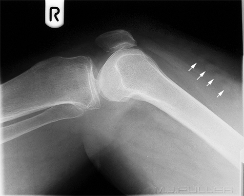
The arrowed soft tissue structure was an incidental finding in a trauma patient.
The arrowed soft tissue structure may be a popliteal artery aneurysm (unproven) The patient's age, sex (male), the site of the lesion and the evidence of atherosclerosis support the diagnosis.
"Popliteal artery aneurysms are the most common peripheral artery aneurysms, comprising 70% to 85% of the total aneurysms in the periphery.1-3 More than 95% of peripheral artery aneurysms occur in males, and the average age of patients at presentation is 65 years. Atherosclerosis appears to be the etiology in more than 90% of cases. The true pathogenesis behind popliteal artery aneurysm formation is not known, and factors such as turbulence distal to the relative stenosis at the tendinous hiatus of the adductor magnus and repeated flexion at the knee have been postulated; this does not, however, account for the association with aneurysms in other locations or the male preponderance. Most popliteal artery aneurysms are fusiform and are bilateral in 25% to 70% of cases." <a class="external" href="http://www.evtoday.com/AAA/2003+Files/Popliteal+Artery+Aneurysms.html" rel="nofollow" target="_blank">Source http://www.evtoday.com/AAA/2003%20Files/Popliteal%20Artery%20Aneurysms.html</a>
