Judet Views

From wikiRadiography
Jump to navigation Jump to search
Introduction

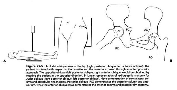
Judet's views are standard radiographic projections which are employed in patients with acetabulum fractures. This page is dedicated to all aspects of Judet view technique.

Radiography
Judet Views
Source:John J. Callaghan, Aaron G. Rosenberg, Harry E. RubashThe adult hip, Volume 1http://books.google.com.au/books?id=CSaFS5Tod3QC&pg=PA360&dq=lateral+hip+radiography&cd=9#v=onepage&q=lateral%20hip%20radiography&f=false
Judet's views are generally only performed as a supplementary view. In cases of acute injury, they can be useful in demonstrating or confirming acetabular fractures. In a clinic referral situation, Judet views may be requested to follow-up fracture healing.

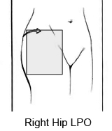
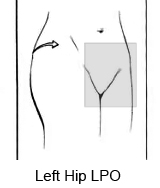
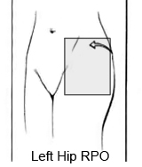
Judet views are basically 45 degree obliques of the affected hip. The 45 degree angle is best achieved by rolling the patient. Alternatively, tube angulation (with careful consideration of grid line orientation) is a legitimate alternative. In patients with acute injuries, rolling the patient will be very painful. Consultation directly with the referring doctor (or preferably the orthopaedic surgeon) will ensure that your approach to performing these views is safe for the patient.

If the Judet views are performed using a bedside technique, a non-grid technique will ensure that the image is not marred by grid cut-off. The image quality which can be achieved with this technique is limited due to excessive scatter radiation. Transfer of the patient onto the X-ray table should be performed with the consent of the referring doctor (and the patient).
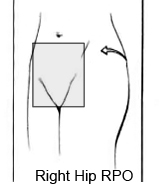
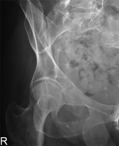
Judet ViewJudet Right Hip RPOThis is the RPO Judet projection of the right hip (left side raised). Note that the patient's right obturator foramen "closes" in this position.
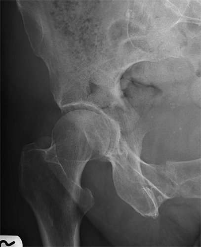
Judet ViewJudet LPO right hipThis is the LPO Judet projection of the right hip. Note that the obturator foramen is "opened" in the position.


Case 1
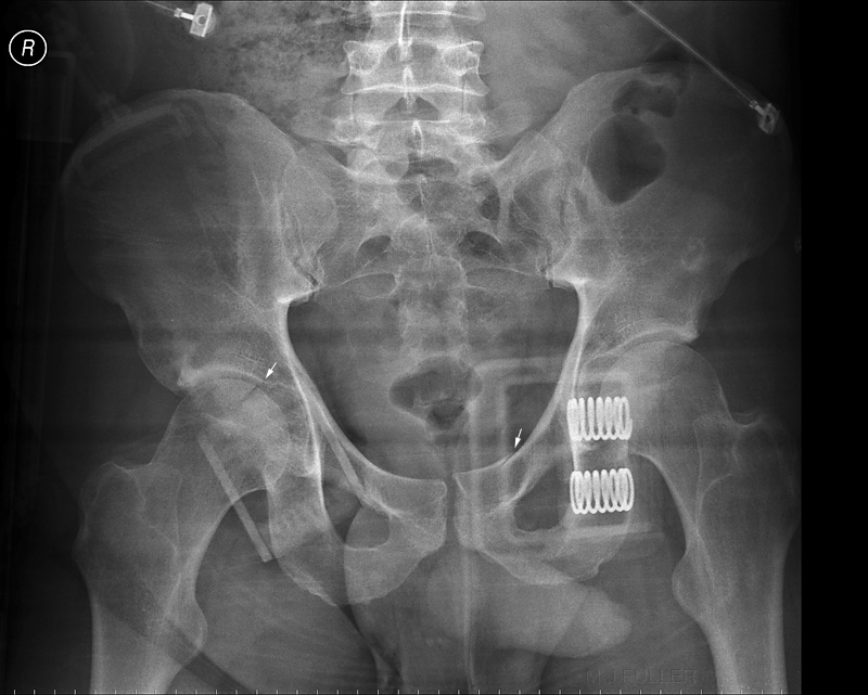
AP PELVISThis is an AP pelvis image which was taken as a part of a trauma series on a patient who presented to the Emergency Department after falling from a height of 4 metres.

There is evidence of an acetabular fracture on the right (arrowed) and a fracture of the superior pubic ramus on the left (arrowed).

The two springs are a part of the compression device that is applied to patients with 'open book' fractures.

The two horizontal dark bands are a known artifacts associated with the joins in the mattress on the resus trolley.
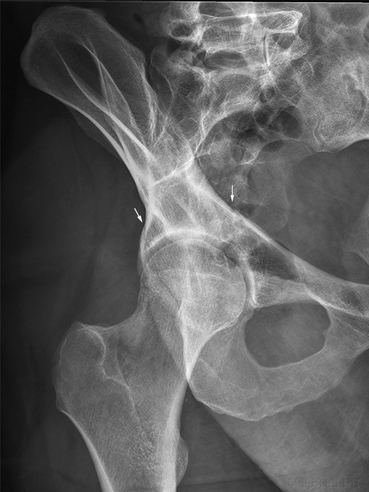
judet viewJudet LPO right hipThe patient was subsequently referred by the orthopaedic surgeon for Judet's views. This is the LPO (right side raised) Judet view. Note that the right SI joint is profiled in this view and the right obturator foramen is 'opened'.

After some deliberation, it was decided to transfer the patient onto the X-ray table. A bedside technique was considered to be likely to produce images of an inadequate quality.

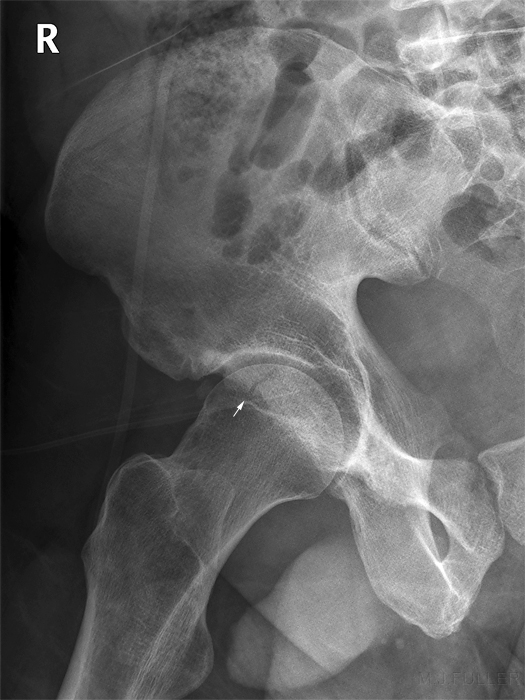
The fracture was demonstrated to extend into the superior margin of the acetabulum and possibly medially into the true pelvis.
judet viewJudet Right Hip RPOThis is the RPO Judet view (patient supine with left side raised to 45 degrees). Note that the obturator foramen is 'closed' and the iliac wing is demonstrated en-face.


Case 2
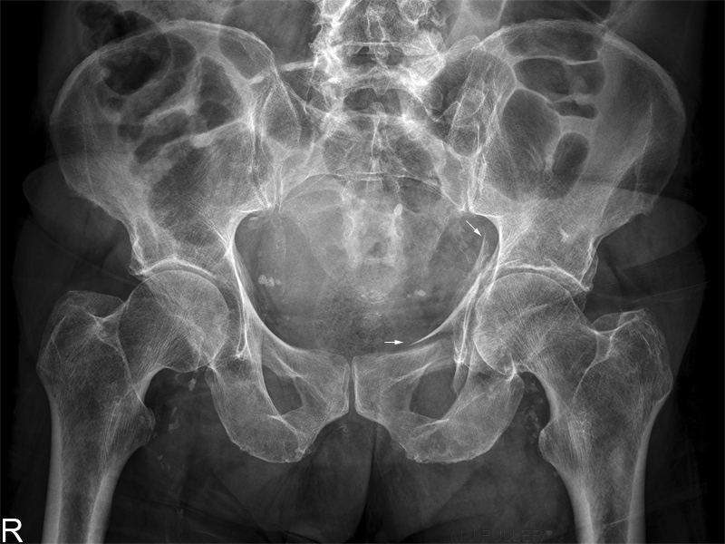
AP PELVISThis 82 year old male presented to the Emergency Department following a fall onto his left side. He was examined and subsequently referred for left hip radiography.
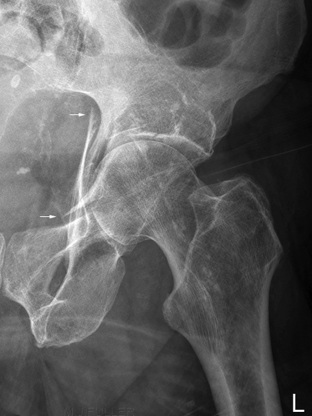
LPO judet viewJudet Left Hip LPOThe LPO Judet View image demonstrates the acetabulum fracture.
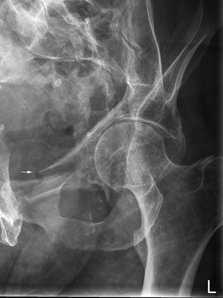
RPO Judet ViewJudet left hip RPOThe RPO Judet view demonstrates the inferior component of the acetabulum fracture


Case 3
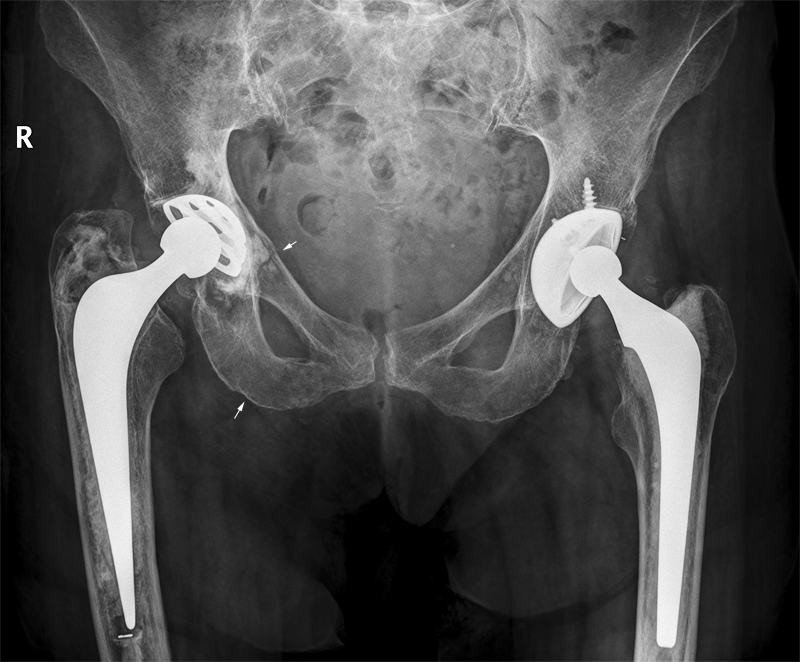
AP pelvisThis 88 year old lady presented to the Emergency Department following a fall at the nursing home. She was examined and referred for radiography of her right hip. The provisional diagnosis was right neck of femur fracture. The referring doctor noted the right leg to be shortened and rotated.

It is not possible to fracture anatomy that was removed surgically several years previously. If the referring doctor had examined the patient he/she would have noticed a surgical scar along the lateral aspect of the proximal femur. It is also noteworthy that it is possible for patients to appear to have a shortened and rotated leg as a result of hip/knee position only.

The AP pelvis image suggested very subtle superior and inferior pubic rami fractures.

The radiographer proceeded to perform Judet views (pubic rami views would also have been a legitimate supplementary view choice)

The patient has bilateral hip replacement prosthesises (the term prosthesii, whilst arguably correct, is not commonly used)
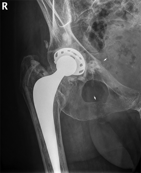
judet viewJudet LPO right hipThe step in the bony contour of the true pelvis is likely to represent an minimally displaced fracture (given the more convincing fracture of the right inferior pubic ramus). This LPO projection of the right hip is a potentially useful choice given the demonstration of the acetabulum and the obturator foramen.
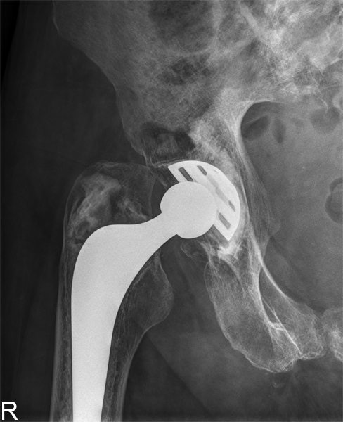
judet viewJudet Right Hip RPOThe RPO Judet view of the right hip did not demonstrate the fractures.


Case 4

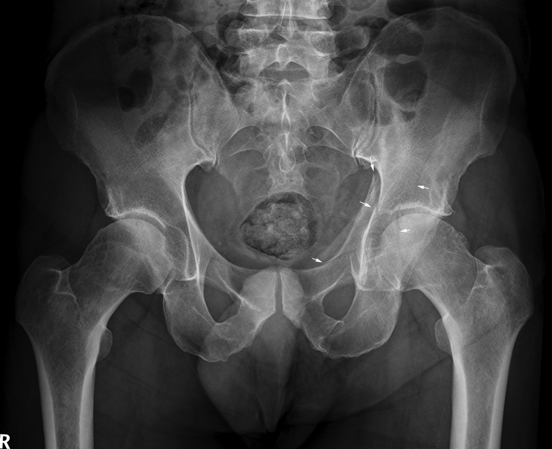
AP PelvisThis 56 year old male presented to the Emergency Department after falling from a ladder. He was experiencing considerable left hip pain and was unable to weightbear. He was referred for left hip radiography.

There is an extensive fracture of the left acetabulum (arrowed).

Note the remarkable similarity with case 2 above.
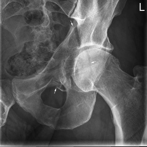
Judet ViewJudet left hip RPOThe radiographer proceeded to perform Judet views to further establish the extent of the fracture.
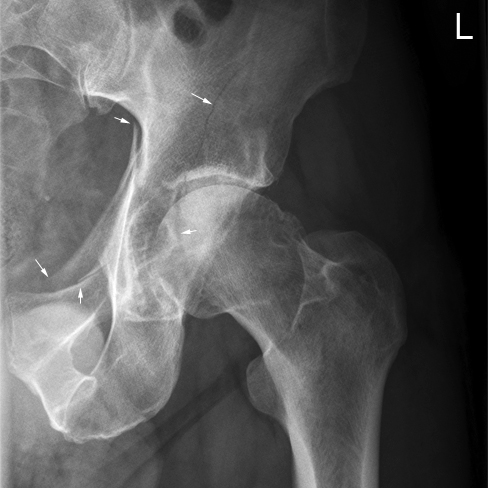
Judet ViewJudet Left Hip LPOThe LPO Judet view demonstrates extension of the fracture into the ilium.

... back to the Wikiradiography Home page
... back to the Applied Radiography home page